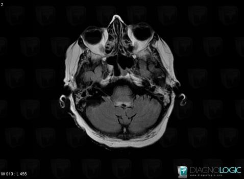
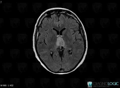
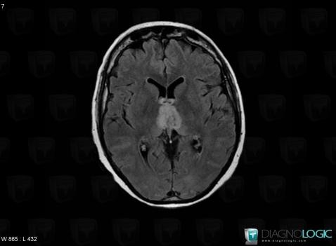
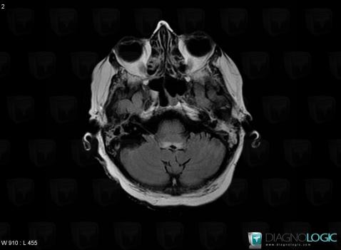
The images below illustrate this case for diagnoses Wernicke encephalopathy, for the modalities (CT ,MRI)
Gamuts for this case : :
- Periventricular anomaly seen in MRI
- Brainstem Hyperintense T2WI or FLAIR lesion
- Brainstem lesion
- Infratentorial T2W or FLAIR hyperintense lesion
- Basal ganglia T2W or FLAIR hyperintense lesion

Here is the specific information in the key image above:
- Diagnosis Wernicke encephalopathy, Location(s) V4 and vermis, with gamuts Periventricular anomaly seen in MRIBrainstem, with gamuts Brainstem Hyperintense T2WI or FLAIR lesion, Brainstem lesionPosterior fossa, with gamuts Infratentorial T2W or FLAIR hyperintense lesion

Here is the specific information in the key image above:
- Diagnosis Wernicke encephalopathy, Location(s) Ventricles / Periventricular region, with gamuts Periventricular anomaly seen in MRIBasal ganglia and capsule, with gamuts Basal ganglia T2W or FLAIR hyperintense lesion

Here is the specific information in the key image above:
- Diagnosis Wernicke encephalopathy, Location(s) Ventricles / Periventricular region, with gamuts Periventricular anomaly seen in MRIBasal ganglia and capsule, with gamuts Basal ganglia T2W or FLAIR hyperintense lesion

Here is the specific information in the key image above:
- Diagnosis Wernicke encephalopathy, Location(s) V4 and vermis, with gamuts Periventricular anomaly seen in MRIBrainstem, with gamuts Brainstem Hyperintense T2WI or FLAIR lesion, Brainstem lesionPosterior fossa, with gamuts Infratentorial T2W or FLAIR hyperintense lesion