The images below illustrate this case for diagnoses Tuberculosis, for the modalities (CT)
Gamuts for this case : :
- Low attenuation mediastinal mass
- Middle mediastinal mass
- Hilar lymph nodes enlargement
- Cystic mediastinal mass
- Localized consolidation
- Large symetric area of ground glass opacity
- Acute consolidation
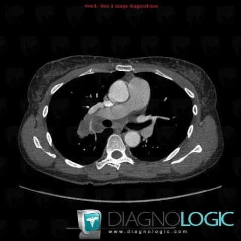
Here is the specific information in the key image above:
- Diagnosis Tuberculosis, Location(s) Mediastinum, with gamuts Middle mediastinal mass
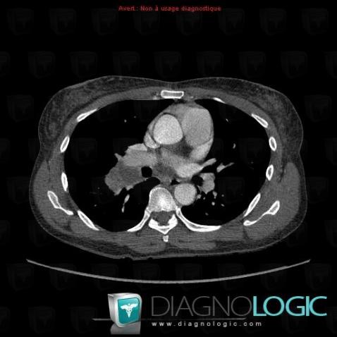
Here is the specific information in the key image above:
- Diagnosis Tuberculosis, Location(s) Mediastinum, with gamuts Hilar lymph nodes enlargement, Cystic mediastinal mass
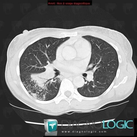
Here is the specific information in the key image above:
- Diagnosis Tuberculosis, Location(s) Pulmonary parenchyma, with gamuts Localized consolidation
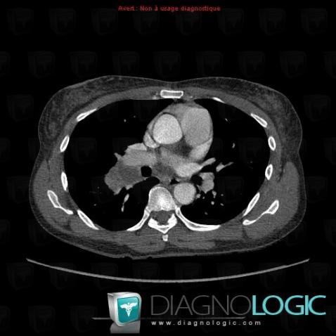
Here is the specific information in the key image above:
- Diagnosis Tuberculosis, Location(s) Mediastinum, with gamuts Hilar lymph nodes enlargement, Cystic mediastinal mass
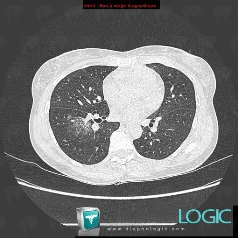
Here is the specific information in the key image above:
- Diagnosis Tuberculosis, Location(s) Pulmonary parenchyma, with gamuts Large symetric area of ground glass opacity
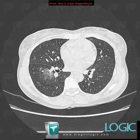
Here is the specific information in the key image above:
- Diagnosis Tuberculosis, Location(s) Pulmonary parenchyma, with gamuts Large symetric area of ground glass opacity
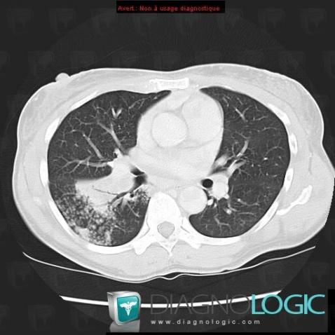
Here is the specific information in the key image above:
- Diagnosis Tuberculosis, Location(s) Pulmonary parenchyma, with gamuts Acute consolidation, Localized consolidation
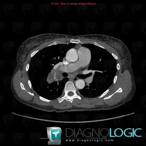
Here is the specific information in the key image above:
- Diagnosis Tuberculosis, Location(s) Mediastinum, with gamuts Low attenuation mediastinal mass, Middle mediastinal mass