The images below illustrate this case for diagnoses Sarcoidosis, for the modalities (CT)
Gamuts for this case : :
- Multiple pulmonary nodule
- Sub pleural pulmonary nodule
- Middle mediastinal mass
- Mediastinal mass with calcifications
- Upper lung zone disease with calcified adenopathy
- Chronic consolidation
- Localized consolidation
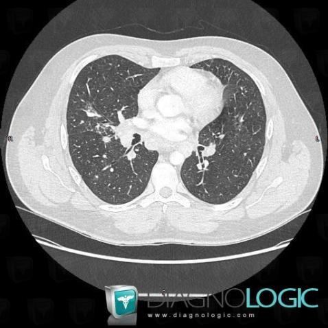
Here is the specific information in the key image above:
- Diagnosis Sarcoidosis, Location(s) Pulmonary parenchyma, with gamuts Multiple pulmonary nodule
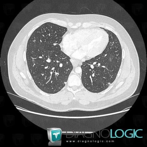
Here is the specific information in the key image above:
- Diagnosis Sarcoidosis, Location(s) Pulmonary parenchyma, with gamuts Sub pleural pulmonary nodule
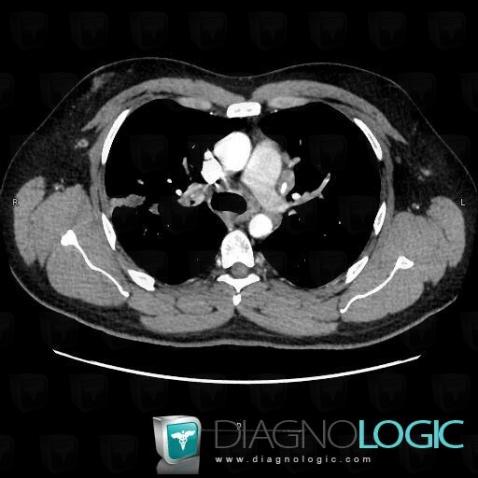
Here is the specific information in the key image above:
- Diagnosis Sarcoidosis, Location(s) Mediastinum, with gamuts Middle mediastinal mass
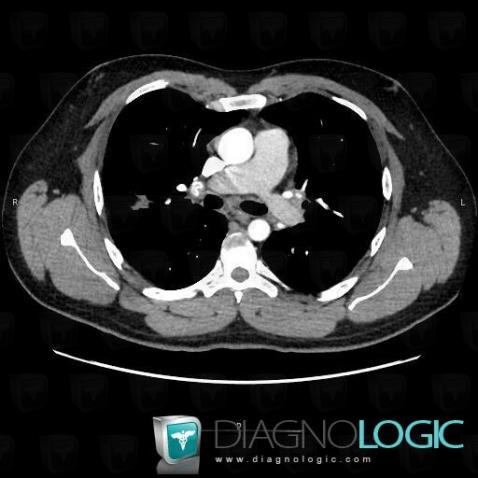
Here is the specific information in the key image above:
- Diagnosis Sarcoidosis, Location(s) Mediastinum, with gamuts Mediastinal mass with calcifications
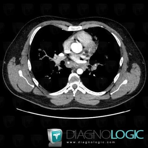
Here is the specific information in the key image above:
- Diagnosis Sarcoidosis, Location(s) Mediastinum, with gamuts Upper lung zone disease with calcified adenopathy
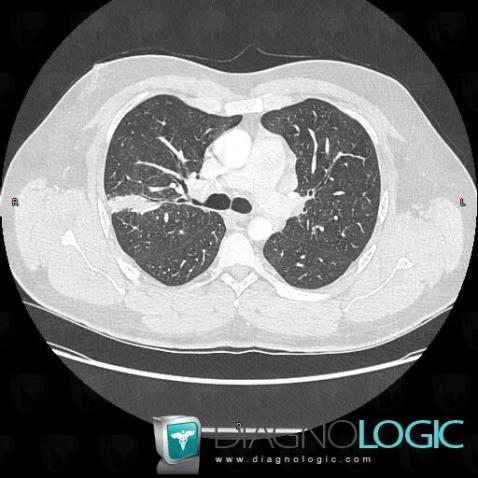
Here is the specific information in the key image above:
- Diagnosis Sarcoidosis, Location(s) Pulmonary parenchyma, with gamuts Chronic consolidation, Localized consolidation



 Patient details - Key images