The images below illustrate this case for diagnoses Sarcoidosis, for the modalities (CT)
Gamuts for this case : :
- Superior mediastinal mass
- Anterior mediastinal mass
- Hilar lymph nodes enlargement
- Mediastinal mass with calcifications
- Irregular peribronchovacular / interlobular thickening
- Perihilar zone disease
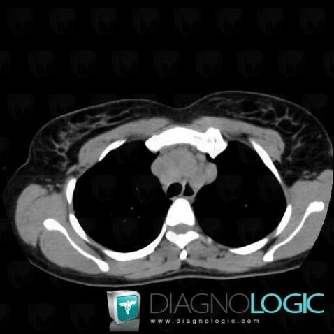
Here is the specific information in the key image above:
- Diagnosis Sarcoidosis, Location(s) Mediastinum, with gamuts Superior mediastinal mass, Anterior mediastinal mass
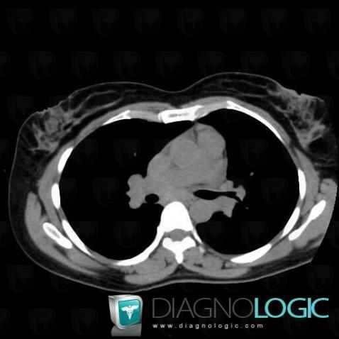
Here is the specific information in the key image above:
- Diagnosis Sarcoidosis, Location(s) Mediastinum, with gamuts Hilar lymph nodes enlargement
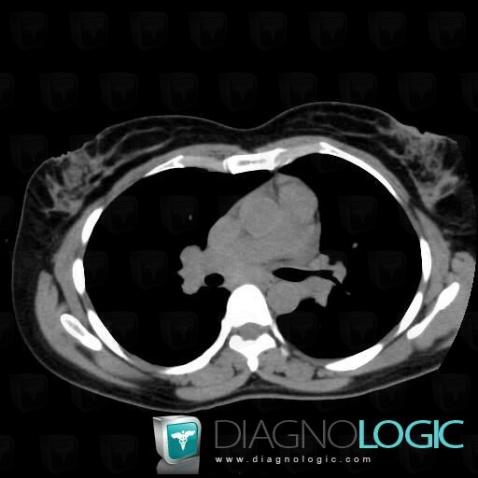
Here is the specific information in the key image above:
- Diagnosis Sarcoidosis, Location(s) Mediastinum, with gamuts Hilar lymph nodes enlargement
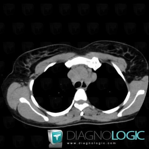
Here is the specific information in the key image above:
- Diagnosis Sarcoidosis, Location(s) Mediastinum, with gamuts Superior mediastinal mass, Anterior mediastinal mass
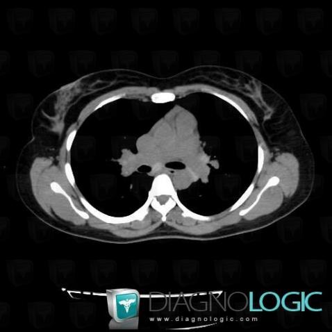
Here is the specific information in the key image above:
- Diagnosis Sarcoidosis, Location(s) Mediastinum, with gamuts Mediastinal mass with calcifications
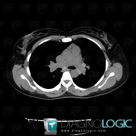
Here is the specific information in the key image above:
- Diagnosis Sarcoidosis, Location(s) Mediastinum, with gamuts Mediastinal mass with calcifications
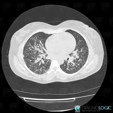
Here is the specific information in the key image above:
- Diagnosis Sarcoidosis, Location(s) Pulmonary parenchyma, with gamuts Irregular peribronchovacular / interlobular thickening
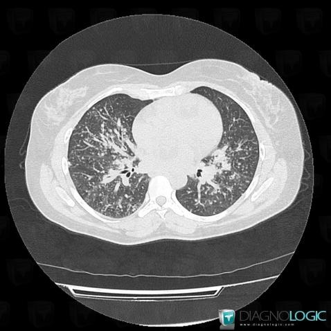
Here is the specific information in the key image above:
- Diagnosis Sarcoidosis, Location(s) Pulmonary parenchyma, with gamuts Irregular peribronchovacular / interlobular thickening
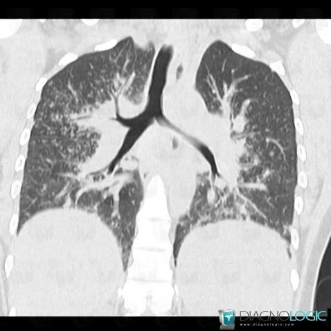
Here is the specific information in the key image above:
- Diagnosis Sarcoidosis, Location(s) Pulmonary parenchyma, with gamuts Perihilar zone disease
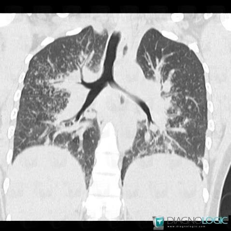
Here is the specific information in the key image above:
- Diagnosis Sarcoidosis, Location(s) Pulmonary parenchyma, with gamuts Perihilar zone disease