The images below illustrate this case for diagnoses Rickets, for the modalities
Gamuts for this case : :
- Fragmented epiphysis
- Transverse sclerotic lines in metaphysis
- sclerotic lines in metaphysis
- Sub periosteal erosion
- Osteolytic lesion of clavicle
- Femoral head acquired deformation
- Skull vault thinning
- Wormian bones
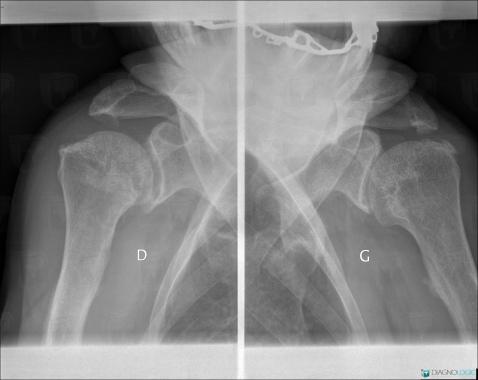
Here is the specific information in the key image above:
- Diagnosis Rickets, Location(s) Femur - Proximal part, with gamuts Fragmented epiphysisScapula, with gamuts Fragmented epiphysisHumerus - Proximal part, with gamuts Fragmented epiphysisHumerus - Distal part, with gamuts Fragmented epiphysisUlna - Proximal part, with gamuts Fragmented epiphysisRadius - Distal part, with gamuts Fragmented epiphysisUlna - Distal part, with gamuts Fragmented epiphysisFemur - Proximal part, with gamuts Fragmented epiphysisFemur - Distal part, with gamuts Fragmented epiphysisTibia - Proximal part, with gamuts Fragmented epiphysisFibula - Proximal part, with gamuts Fragmented epiphysisTibia - Distal part, with gamuts Fragmented epiphysisFibula - Distal part, with gamuts Fragmented epiphysisRadius - Proximal part, with gamuts Fragmented epiphysis
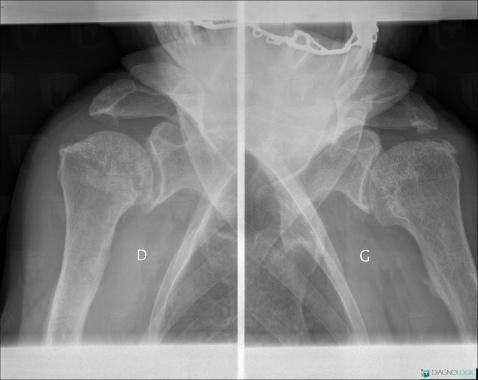
Here is the specific information in the key image above:
- Diagnosis Rickets, Location(s) Humerus - Proximal part, with gamuts Transverse sclerotic lines in metaphysisHumerus - Distal part, with gamuts Transverse sclerotic lines in metaphysisUlna - Proximal part, with gamuts Transverse sclerotic lines in metaphysisRadius - Distal part, with gamuts Transverse sclerotic lines in metaphysisUlna - Distal part, with gamuts Transverse sclerotic lines in metaphysisFemur - Proximal part, with gamuts Transverse sclerotic lines in metaphysisFemur - Distal part, with gamuts Transverse sclerotic lines in metaphysisTibia - Proximal part, with gamuts Transverse sclerotic lines in metaphysisFibula - Proximal part, with gamuts Transverse sclerotic lines in metaphysisTibia - Distal part, with gamuts Transverse sclerotic lines in metaphysisFibula - Distal part, with gamuts Transverse sclerotic lines in metaphysisRadius - Proximal part, with gamuts Transverse sclerotic lines in metaphysis
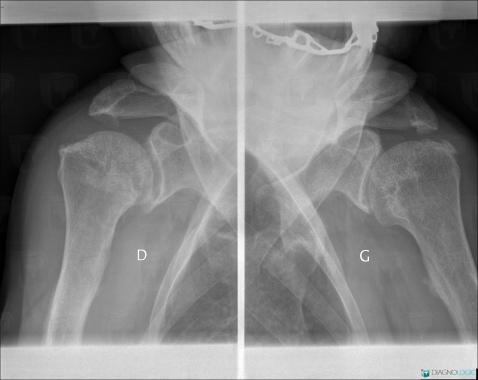
Here is the specific information in the key image above:
- Diagnosis Rickets, Location(s) Scapula, with gamuts sclerotic lines in metaphysisHumerus - Proximal part, with gamuts sclerotic lines in metaphysisHumerus - Distal part, with gamuts sclerotic lines in metaphysisUlna - Proximal part, with gamuts sclerotic lines in metaphysisRadius - Distal part, with gamuts sclerotic lines in metaphysisUlna - Distal part, with gamuts sclerotic lines in metaphysisFemur - Proximal part, with gamuts sclerotic lines in metaphysisFemur - Distal part, with gamuts sclerotic lines in metaphysisTibia - Proximal part, with gamuts sclerotic lines in metaphysisFibula - Proximal part, with gamuts sclerotic lines in metaphysisTibia - Distal part, with gamuts sclerotic lines in metaphysisFibula - Distal part, with gamuts sclerotic lines in metaphysisRadius - Proximal part, with gamuts sclerotic lines in metaphysis
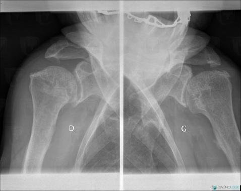
Here is the specific information in the key image above:
- Diagnosis Rickets, Location(s) Scapula, with gamuts Humerus - Proximal part, with gamuts Humerus - Distal part, with gamuts Ulna - Proximal part, with gamuts Radius - Distal part, with gamuts Ulna - Distal part, with gamuts Femur - Proximal part, with gamuts Femur - Distal part, with gamuts Tibia - Proximal part, with gamuts Fibula - Proximal part, with gamuts Tibia - Distal part, with gamuts Fibula - Distal part, with gamuts Radius - Proximal part, with gamuts
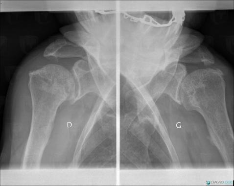
Here is the specific information in the key image above:
- Diagnosis Rickets, Location(s) Scapula, with gamuts Sub periosteal erosionHumerus - Proximal part, with gamuts Sub periosteal erosionHumerus - Mid part, with gamuts Sub periosteal erosionHumerus - Distal part, with gamuts Sub periosteal erosionUlna - Proximal part, with gamuts Sub periosteal erosionRadius - Mid part, with gamuts Sub periosteal erosionUlna - Mid part, with gamuts Sub periosteal erosionRadius - Distal part, with gamuts Sub periosteal erosionUlna - Distal part, with gamuts Sub periosteal erosionFemur - Proximal part, with gamuts Sub periosteal erosionFemur - Mid part, with gamuts Sub periosteal erosionFemur - Distal part, with gamuts Sub periosteal erosionTibia - Proximal part, with gamuts Sub periosteal erosionFibula - Proximal part, with gamuts Sub periosteal erosionTibia - Mid part, with gamuts Sub periosteal erosionFibula - Mid part, with gamuts Sub periosteal erosionTibia - Distal part, with gamuts Sub periosteal erosionFibula - Distal part, with gamuts Sub periosteal erosionRadius - Proximal part, with gamuts Sub periosteal erosion
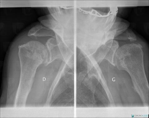
Here is the specific information in the key image above:
- Diagnosis Rickets, Location(s) Clavicle, with gamuts Osteolytic lesion of clavicle
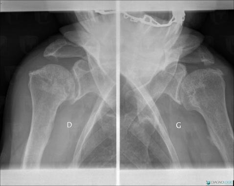
Here is the specific information in the key image above:
- Diagnosis Rickets, Location(s) Femur - Proximal part, with gamuts Femoral head acquired deformationFemur - Proximal part, with gamuts Femoral head acquired deformation
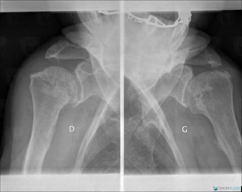
Here is the specific information in the key image above:
- Diagnosis Rickets, Location(s) Skull vault, with gamuts Skull vault thinningSkull vault, with gamuts Skull vault thinning
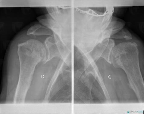
Here is the specific information in the key image above:
- Diagnosis Rickets, Location(s) Skull vault, with gamuts Wormian bonesSkull vault, with gamuts Wormian bones