The images below illustrate this case for diagnoses Radiation pneumonitis, Venous compression, for the modalities (CT)
Gamuts for this case : :
- Upper lung zone disease
- Pulmonary fibrosis
- Chronic consolidation
- Localized consolidation
- Smooth peribronchovacular / interlobular thickening
- Peribronchovacular / interlobular thickening
- thoracic vascular disease
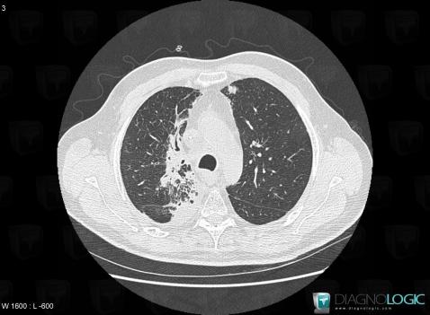
Here is the specific information in the key image above:
- Diagnosis Radiation pneumonitis, Location(s) Pulmonary parenchyma, with gamuts Upper lung zone disease, Pulmonary fibrosis, Chronic consolidation, Localized consolidation
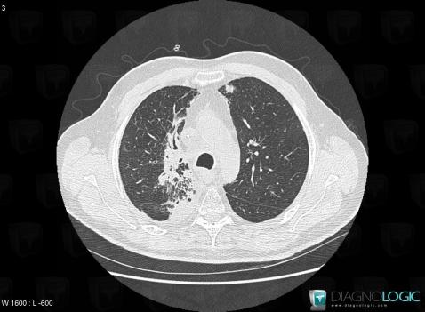
Here is the specific information in the key image above:
- Diagnosis Radiation pneumonitis, Location(s) Pulmonary parenchyma, with gamuts Upper lung zone disease, Pulmonary fibrosis, Chronic consolidation, Localized consolidation
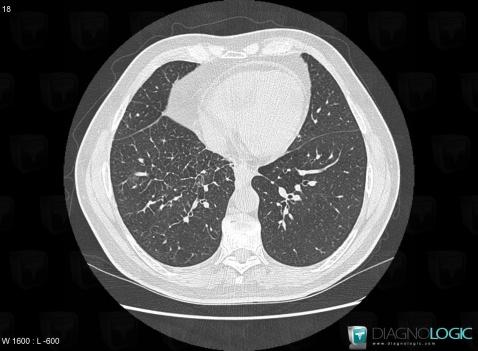
Here is the specific information in the key image above:
- Diagnosis Venous compression, Location(s) Pulmonary parenchyma, with gamuts Smooth peribronchovacular / interlobular thickening, Peribronchovacular / interlobular thickening
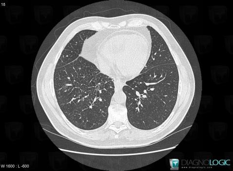
Here is the specific information in the key image above:
- Diagnosis Venous compression, Location(s) Pulmonary parenchyma, with gamuts Smooth peribronchovacular / interlobular thickening, Peribronchovacular / interlobular thickening
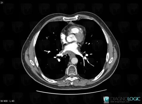
Here is the specific information in the key image above:
- Diagnosis Venous compression, Location(s) Veins - Thorax, with gamuts thoracic vascular disease
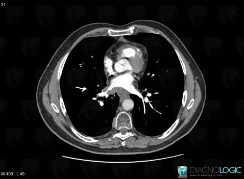
Here is the specific information in the key image above:
- Diagnosis Venous compression, Location(s) Veins - Thorax, with gamuts thoracic vascular disease