The images below illustrate this case for diagnoses Pulmonary infarction, Pulmonary thromboembolism, for the modalities (CT)
Gamuts for this case : :
- thoracic vascular disease
- Peripheral and sub pleural zone disease
- Large symetric area of ground glass opacity
- Sub pleural pulmonary nodule
- Acute consolidation
- Localized consolidation
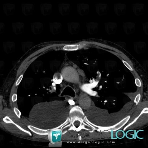
Here is the specific information in the key image above:
- Diagnosis Pulmonary thromboembolism, Location(s) Pulmonary artery, with gamuts thoracic vascular disease
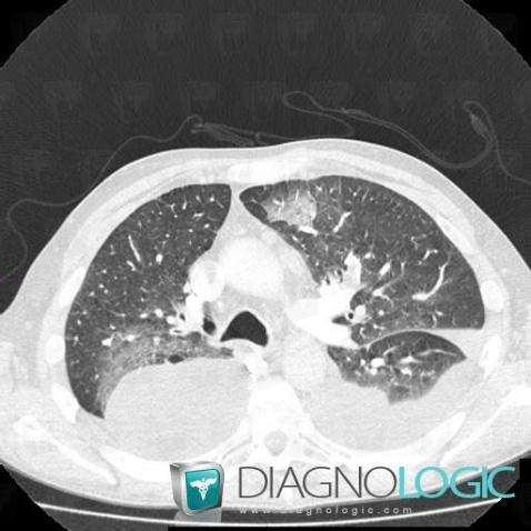
Here is the specific information in the key image above:
- Diagnosis Pulmonary infarction, Location(s) Pulmonary parenchyma, with gamuts Peripheral and sub pleural zone disease, Large symetric area of ground glass opacity
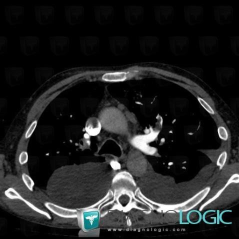
Here is the specific information in the key image above:
- Diagnosis Pulmonary thromboembolism, Location(s) Pulmonary artery, with gamuts thoracic vascular disease
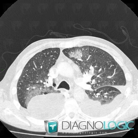
Here is the specific information in the key image above:
- Diagnosis Pulmonary infarction, Location(s) Pulmonary parenchyma, with gamuts Peripheral and sub pleural zone disease, Large symetric area of ground glass opacity
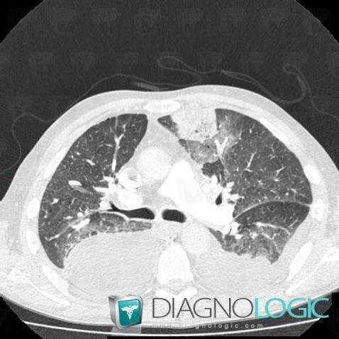
Here is the specific information in the key image above:
- Diagnosis Pulmonary infarction, Location(s) Pulmonary parenchyma, with gamuts Sub pleural pulmonary nodule, Acute consolidation, Peripheral and sub pleural zone disease, Localized consolidation
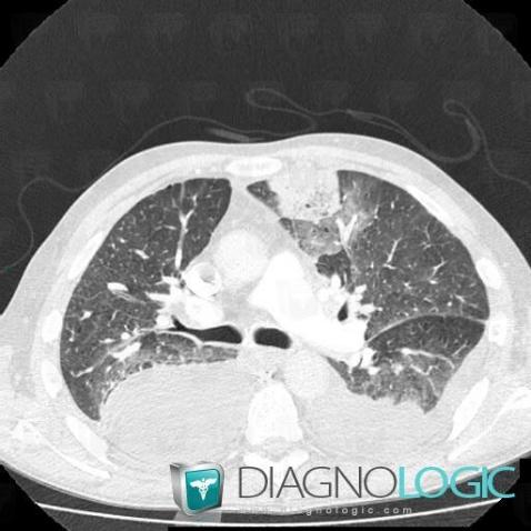
Here is the specific information in the key image above:
- Diagnosis Pulmonary infarction, Location(s) Pulmonary parenchyma, with gamuts Sub pleural pulmonary nodule, Acute consolidation, Peripheral and sub pleural zone disease, Localized consolidation