The images below illustrate this case for diagnoses Sarcoidosis (link to Pulmonary fibrosis), Sarcoidosis, for the modalities (CT)
Gamuts for this case : :
- Pleural thickening
- Irregular peribronchovacular / interlobular thickening
- Multiple pulmonary nodule
- Nodular peribronchovacular / interlobular thickening
- Perihilar zone disease
- Peripheral and sub pleural zone disease
- Middle mediastinal mass
- Upper lung zone disease with calcified adenopathy
- Mediastinal mass with calcifications
- Hilar lymph nodes enlargement
- Posterior mediastinal mass
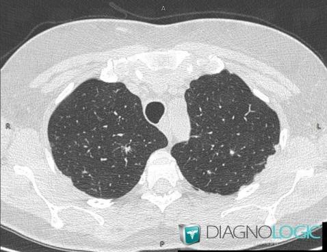
Here is the specific information in the key image above:
- Diagnosis Sarcoidosis, Location(s) Pleura, with gamuts Pleural thickening
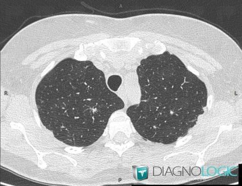
Here is the specific information in the key image above:
- Diagnosis Sarcoidosis, Location(s) Pleura, with gamuts Pleural thickening
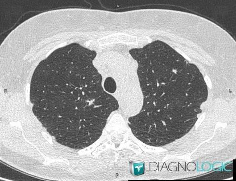
Here is the specific information in the key image above:
- Diagnosis Sarcoidosis, Location(s) Pulmonary parenchyma, with gamuts Irregular peribronchovacular / interlobular thickening
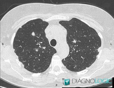
Here is the specific information in the key image above:
- Diagnosis Sarcoidosis, Location(s) Pulmonary parenchyma, with gamuts Multiple pulmonary nodule
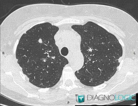
Here is the specific information in the key image above:
- Diagnosis Sarcoidosis, Location(s) Pulmonary parenchyma, with gamuts Multiple pulmonary nodule
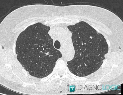
Here is the specific information in the key image above:
- Diagnosis Sarcoidosis, Location(s) Pulmonary parenchyma, with gamuts Irregular peribronchovacular / interlobular thickening
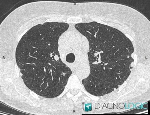
Here is the specific information in the key image above:
- Diagnosis Sarcoidosis (link to Pulmonary fibrosis), Location(s) Pulmonary parenchyma, with gamuts Nodular peribronchovacular / interlobular thickening
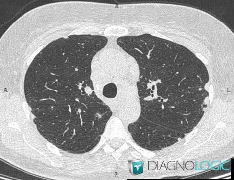
Here is the specific information in the key image above:
- Diagnosis Sarcoidosis (link to Pulmonary fibrosis), Location(s) Pulmonary parenchyma, with gamuts Nodular peribronchovacular / interlobular thickening
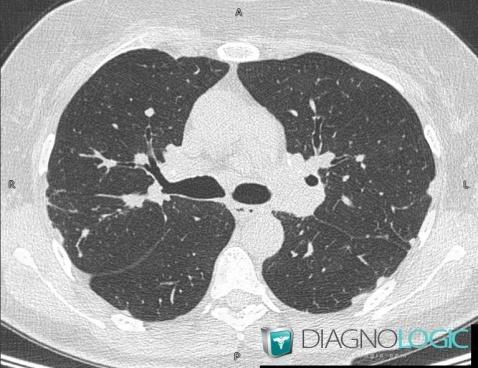
Here is the specific information in the key image above:
- Diagnosis Sarcoidosis, Location(s) Pulmonary parenchyma, with gamuts Perihilar zone disease
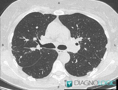
Here is the specific information in the key image above:
- Diagnosis Sarcoidosis, Location(s) Pulmonary parenchyma, with gamuts Perihilar zone disease
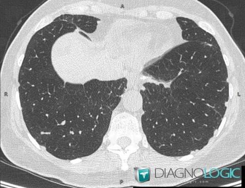
Here is the specific information in the key image above:
- Diagnosis Sarcoidosis, Location(s) Pulmonary parenchyma, with gamuts Peripheral and sub pleural zone disease
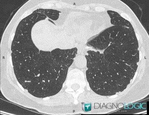
Here is the specific information in the key image above:
- Diagnosis Sarcoidosis, Location(s) Pulmonary parenchyma, with gamuts Peripheral and sub pleural zone disease
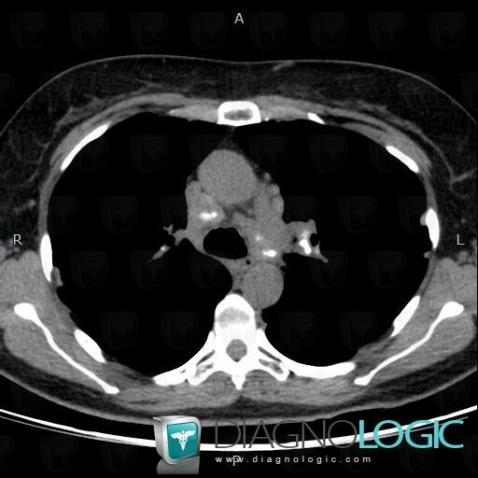
Here is the specific information in the key image above:
- Diagnosis Sarcoidosis, Location(s) Mediastinum, with gamuts Middle mediastinal mass, Upper lung zone disease with calcified adenopathy, Mediastinal mass with calcifications
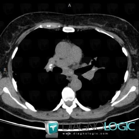
Here is the specific information in the key image above:
- Diagnosis Sarcoidosis, Location(s) Mediastinum, with gamuts Hilar lymph nodes enlargement
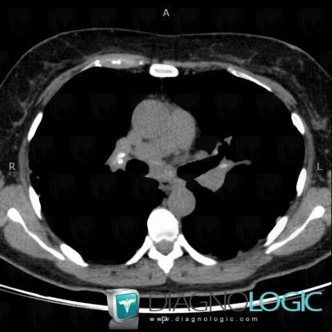
Here is the specific information in the key image above:
- Diagnosis Sarcoidosis, Location(s) Mediastinum, with gamuts Hilar lymph nodes enlargement
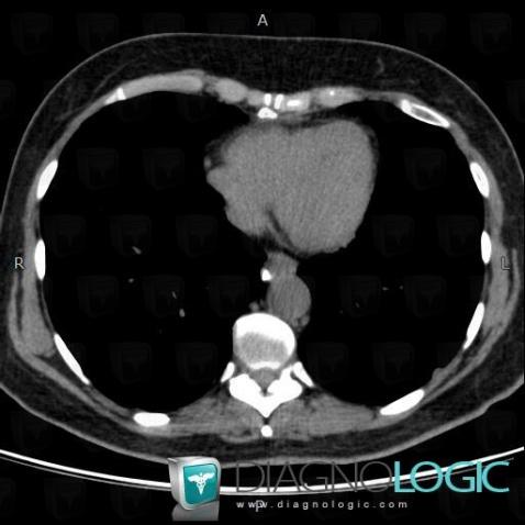
Here is the specific information in the key image above:
- Diagnosis Sarcoidosis, Location(s) Mediastinum, with gamuts Posterior mediastinal mass
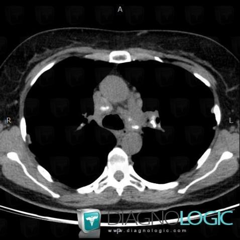
Here is the specific information in the key image above:
- Diagnosis Sarcoidosis, Location(s) Mediastinum, with gamuts Middle mediastinal mass, Upper lung zone disease with calcified adenopathy, Mediastinal mass with calcifications
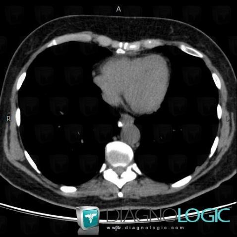
Here is the specific information in the key image above:
- Diagnosis Sarcoidosis, Location(s) Mediastinum, with gamuts Posterior mediastinal mass