The images below illustrate this case for diagnoses Pulmonary edema, Pleural effusion, for the modalities (CT ,X rays)
Gamuts for this case : :
- Pleural calcification
- Large symetric area of ground glass opacity
- Bronchial wall thickening
- Perihilar zone disease
- Pleural thickening
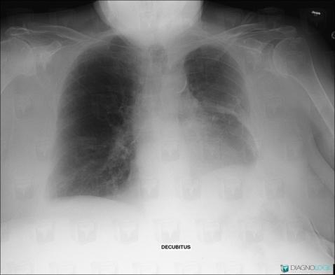
Here is the specific information in the key image above:
- Diagnosis Pleural effusion, Location(s) Pleura, with gamuts Pleural calcification
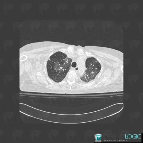
Here is the specific information in the key image above:
- Diagnosis Pulmonary edema, Location(s) Pulmonary parenchyma, with gamuts Large symetric area of ground glass opacity
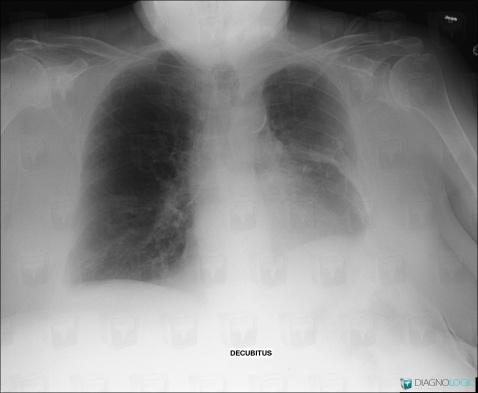
Here is the specific information in the key image above:
- Diagnosis Pleural effusion, Location(s) Pleura, with gamuts Pleural calcification
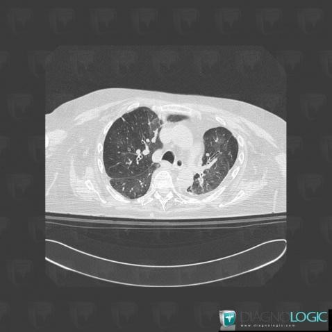
Here is the specific information in the key image above:
- Diagnosis Pulmonary edema, Location(s) Airways, with gamuts Bronchial wall thickening
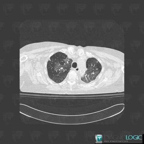
Here is the specific information in the key image above:
- Diagnosis Pulmonary edema, Location(s) Pulmonary parenchyma, with gamuts Large symetric area of ground glass opacity
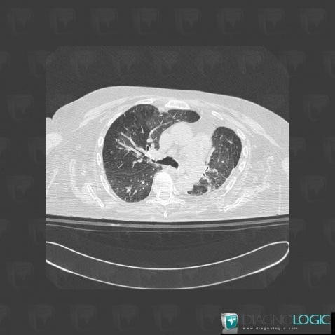
Here is the specific information in the key image above:
- Diagnosis Pulmonary edema, Location(s) Pulmonary parenchyma, with gamuts Perihilar zone disease
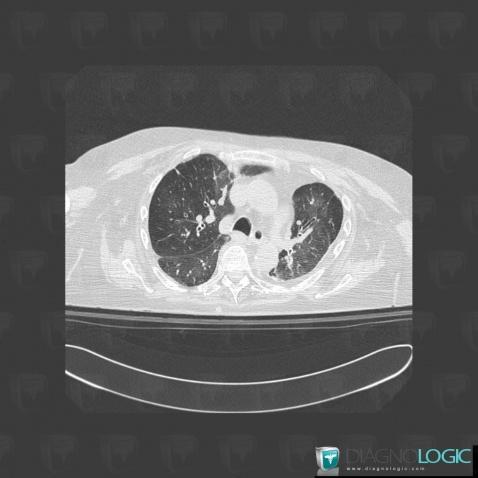
Here is the specific information in the key image above:
- Diagnosis Pulmonary edema, Location(s) Airways, with gamuts Bronchial wall thickening
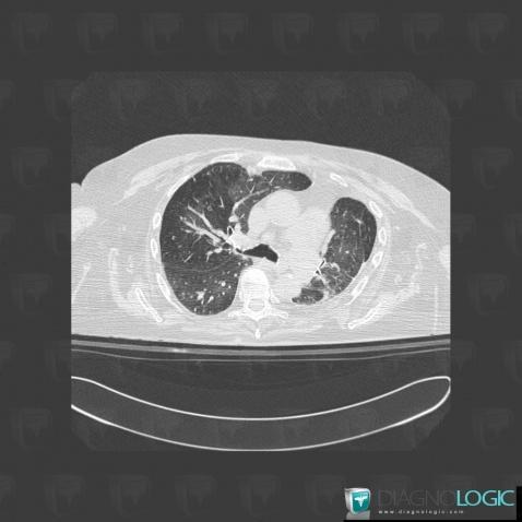
Here is the specific information in the key image above:
- Diagnosis Pulmonary edema, Location(s) Pulmonary parenchyma, with gamuts Perihilar zone disease
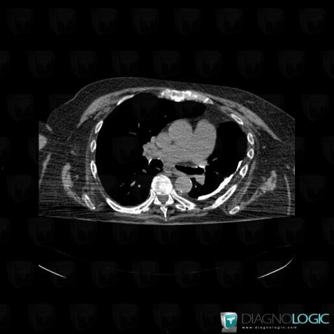
Here is the specific information in the key image above:
- Diagnosis Pleural effusion, Location(s) Pleura, with gamuts Pleural calcification, Pleural thickening
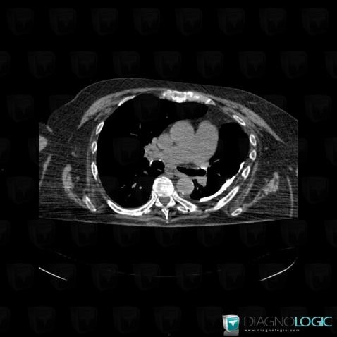
Here is the specific information in the key image above:
- Diagnosis Pleural effusion, Location(s) Pleura, with gamuts Pleural calcification, Pleural thickening