The images below illustrate this case for diagnoses Metastasis (link to Pineal tumor), Metastasis, Bronchogenic carcinoma, for the modalities (MRI ,CT)
Gamuts for this case : :
- Infratentorial T2W or FLAIR hypointense lesion
- Infratentorial T2W or FLAIR hyperintense lesion
- Brainstem Hyperintense T2WI or FLAIR lesion
- Brainstem lesion
- Periventricular anomaly seen in MRI
- Intracerebral T2W or FLAIR hypointense lesion
- Intracerebral T2W or FLAIR hyperintense lesion
- Intracerebral T2W or FLAIR isointense lesion
- Without gamut
- Infratentorial lesion with moderate enhancement
- Infratentorial lesion with ring enhancement
- Multifocal infratentorial lesions
- Cerebellar lesion
- Multifocal intracranial lesions
- Midline mass
- Intracerebral lesion with moderate enhancement
- Intracerebral lesion with ring enhancement
- Superior mediastinal mass
- Mediastinal mass with marked enhancement
- Middle mediastinal mass
- Low density in portal phase hepatic lesion
- Solid hepatic lesion
- Pulmonary mass with hilar attachment
- Solitary pulmonary mass
- Infratentorial cystic mass mass with a mural nodule
- Infratentorial lesion with intense enhancement
- Subcortical lesion
- Intracerebral lesion with intense enhancement
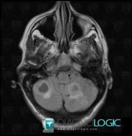
Here is the specific information in the key image above:
- Diagnosis Metastasis, Location(s) Posterior fossa, with gamuts Infratentorial T2W or FLAIR hypointense lesion, Infratentorial T2W or FLAIR hyperintense lesion
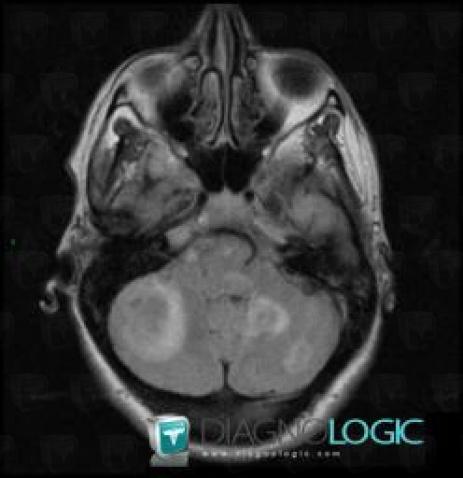
Here is the specific information in the key image above:
- Diagnosis Metastasis, Location(s) Brainstem, with gamuts Brainstem Hyperintense T2WI or FLAIR lesion, Brainstem lesion
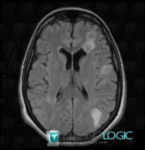
Here is the specific information in the key image above:
- Diagnosis Metastasis, Location(s) Ventricles / Periventricular region, with gamuts Periventricular anomaly seen in MRI
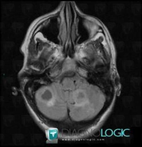
Here is the specific information in the key image above:
- Diagnosis Metastasis, Location(s) Posterior fossa, with gamuts Infratentorial T2W or FLAIR hypointense lesion, Infratentorial T2W or FLAIR hyperintense lesion
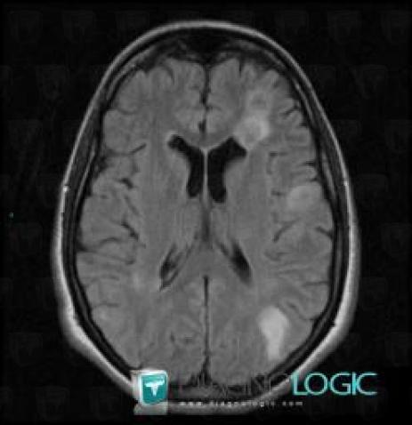
Here is the specific information in the key image above:
- Diagnosis Metastasis, Location(s) Ventricles / Periventricular region, with gamuts Periventricular anomaly seen in MRI
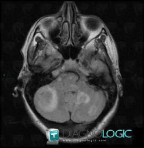
Here is the specific information in the key image above:
- Diagnosis Metastasis, Location(s) Brainstem, with gamuts Brainstem Hyperintense T2WI or FLAIR lesion, Brainstem lesion
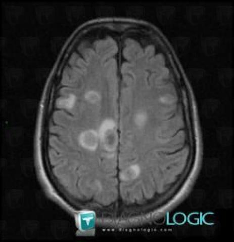
Here is the specific information in the key image above:
- Diagnosis Metastasis, Location(s) Cerebral hemispheres, with gamuts Intracerebral T2W or FLAIR hypointense lesion
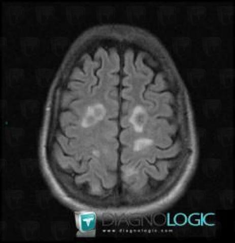
Here is the specific information in the key image above:
- Diagnosis Metastasis, Location(s) Cerebral hemispheres, with gamuts Intracerebral T2W or FLAIR hyperintense lesion, Intracerebral T2W or FLAIR isointense lesion
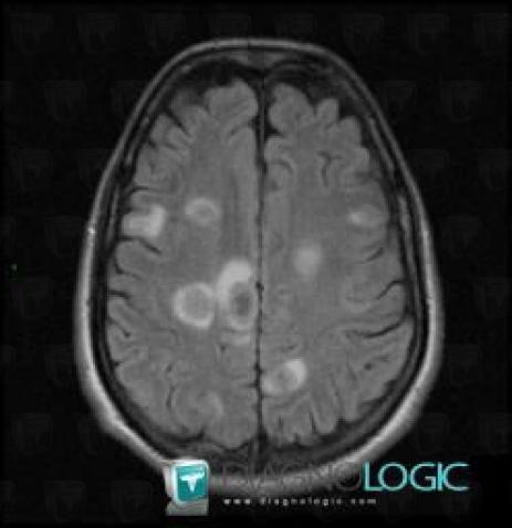
Here is the specific information in the key image above:
- Diagnosis Metastasis, Location(s) Cerebral hemispheres, with gamuts Intracerebral T2W or FLAIR hypointense lesion
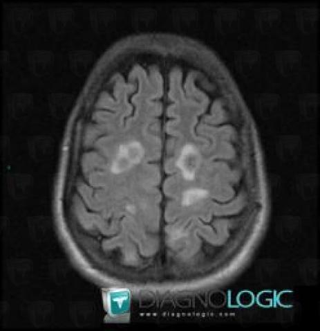
Here is the specific information in the key image above:
- Diagnosis Metastasis, Location(s) Cerebral hemispheres, with gamuts Intracerebral T2W or FLAIR hyperintense lesion, Intracerebral T2W or FLAIR isointense lesion
Here is the specific information in the key image above:
- Diagnosis Metastasis, Location(s) Cerebral hemispheres, with gamuts
Here is the specific information in the key image above:
- Diagnosis Metastasis, Location(s) Cerebral hemispheres, with gamuts
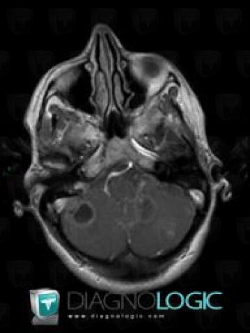
Here is the specific information in the key image above:
- Diagnosis Metastasis, Location(s) Posterior fossa, with gamuts Infratentorial lesion with moderate enhancement, Infratentorial lesion with ring enhancement, Multifocal infratentorial lesions
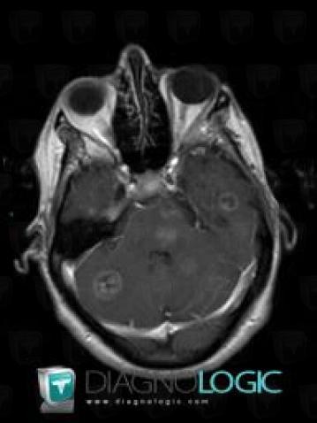
Here is the specific information in the key image above:
- Diagnosis Metastasis, Location(s) Cerebellar hemisphere, with gamuts Cerebellar lesion
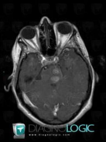
Here is the specific information in the key image above:
- Diagnosis Metastasis, Location(s) Brainstem, with gamuts Brainstem lesion
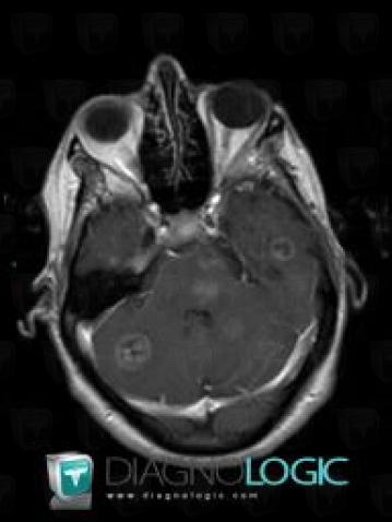
Here is the specific information in the key image above:
- Diagnosis Metastasis, Location(s) Cerebellar hemisphere, with gamuts Cerebellar lesion
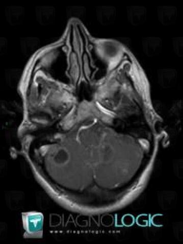
Here is the specific information in the key image above:
- Diagnosis Metastasis, Location(s) Posterior fossa, with gamuts Infratentorial lesion with moderate enhancement, Infratentorial lesion with ring enhancement, Multifocal infratentorial lesions
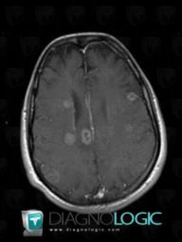
Here is the specific information in the key image above:
- Diagnosis Metastasis, Location(s) Cerebral hemispheres, with gamuts Multifocal intracranial lesions, Intracerebral lesion with moderate enhancement
- Diagnosis Metastasis (link to Pineal tumor), Location(s) Cerebral falx / Midline, with gamuts Midline mass
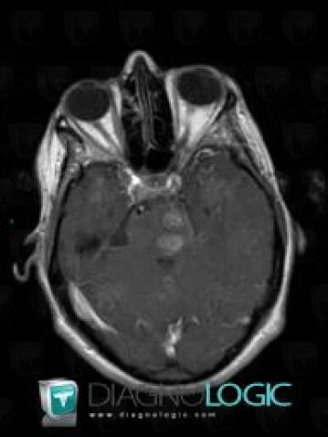
Here is the specific information in the key image above:
- Diagnosis Metastasis, Location(s) Brainstem, with gamuts Brainstem lesion
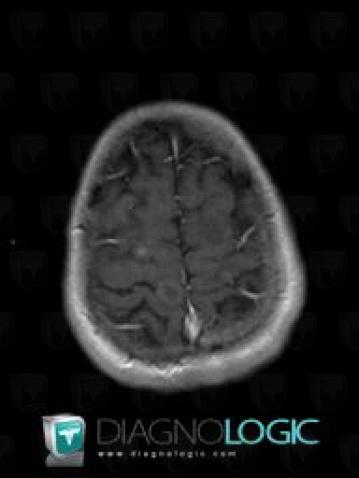
Here is the specific information in the key image above:
- Diagnosis Metastasis, Location(s) Cerebral hemispheres, with gamuts Intracerebral lesion with ring enhancement
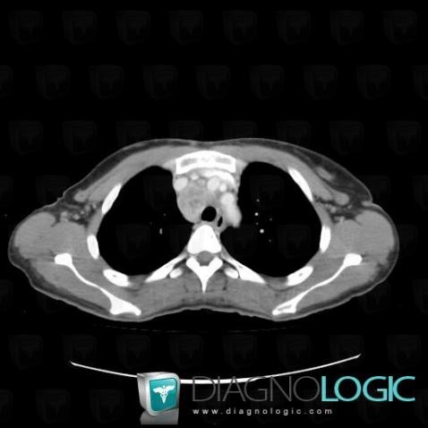
Here is the specific information in the key image above:
- Diagnosis Metastasis, Location(s) Mediastinum, with gamuts Superior mediastinal mass, Mediastinal mass with marked enhancement
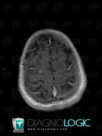
Here is the specific information in the key image above:
- Diagnosis Metastasis, Location(s) Cerebral hemispheres, with gamuts Intracerebral lesion with ring enhancement
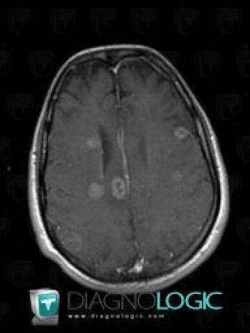
Here is the specific information in the key image above:
- Diagnosis Metastasis, Location(s) Cerebral hemispheres, with gamuts Multifocal intracranial lesions, Intracerebral lesion with moderate enhancement
- Diagnosis Metastasis (link to Pineal tumor), Location(s) Cerebral falx / Midline, with gamuts Midline mass
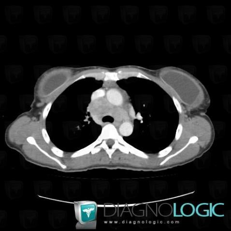
Here is the specific information in the key image above:
- Diagnosis Metastasis, Location(s) Mediastinum, with gamuts Middle mediastinal mass
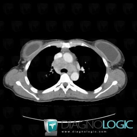
Here is the specific information in the key image above:
- Diagnosis Metastasis, Location(s) Mediastinum, with gamuts Middle mediastinal mass
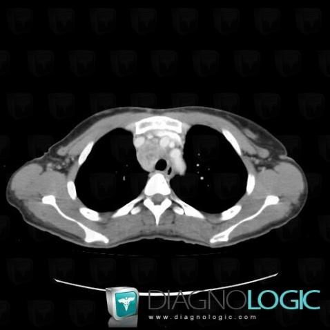
Here is the specific information in the key image above:
- Diagnosis Metastasis, Location(s) Mediastinum, with gamuts Superior mediastinal mass, Mediastinal mass with marked enhancement
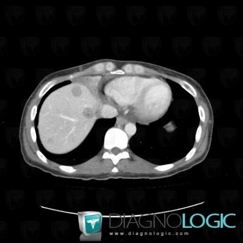
Here is the specific information in the key image above:
- Diagnosis Metastasis, Location(s) Liver, with gamuts Low density in portal phase hepatic lesion, Solid hepatic lesion
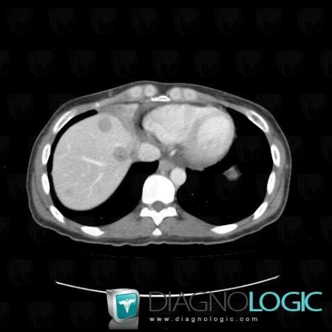
Here is the specific information in the key image above:
- Diagnosis Metastasis, Location(s) Liver, with gamuts Low density in portal phase hepatic lesion, Solid hepatic lesion
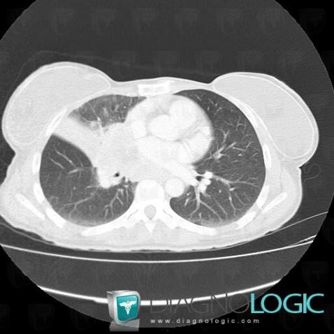
Here is the specific information in the key image above:
- Diagnosis Bronchogenic carcinoma, Location(s) Pulmonary parenchyma, with gamuts Pulmonary mass with hilar attachment, Solitary pulmonary mass
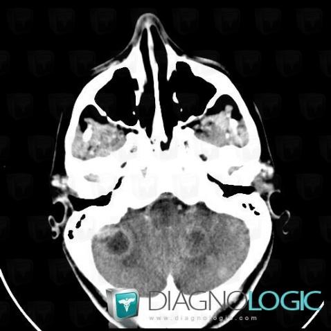
Here is the specific information in the key image above:
- Diagnosis Metastasis, Location(s) Posterior fossa, with gamuts Infratentorial cystic mass mass with a mural nodule, Infratentorial lesion with ring enhancement, Infratentorial lesion with moderate enhancement, Multifocal infratentorial lesionsCerebellar hemisphere, with gamuts Cerebellar lesion
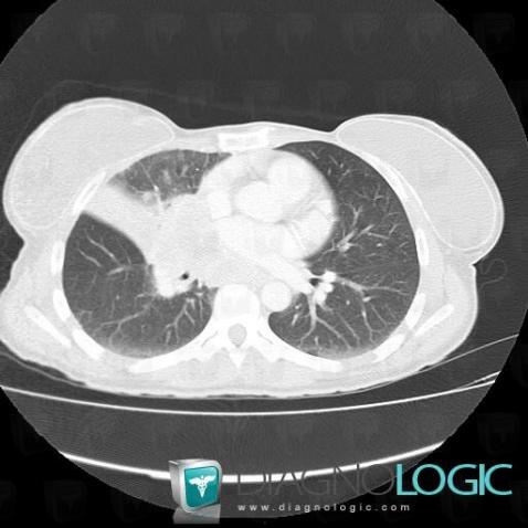
Here is the specific information in the key image above:
- Diagnosis Bronchogenic carcinoma, Location(s) Pulmonary parenchyma, with gamuts Pulmonary mass with hilar attachment, Solitary pulmonary mass
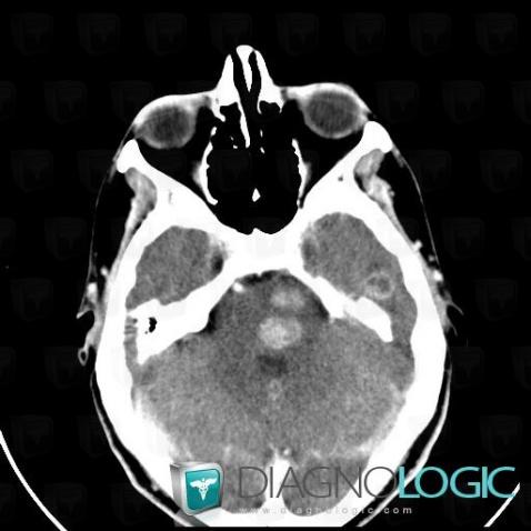
Here is the specific information in the key image above:
- Diagnosis Metastasis, Location(s) Brainstem, with gamuts Brainstem lesion
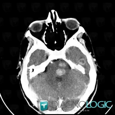
Here is the specific information in the key image above:
- Diagnosis Metastasis, Location(s) Brainstem, with gamuts Brainstem lesion
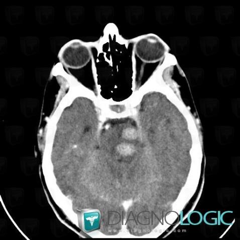
Here is the specific information in the key image above:
- Diagnosis Metastasis, Location(s) Posterior fossa, with gamuts Infratentorial lesion with intense enhancement
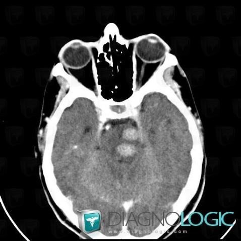
Here is the specific information in the key image above:
- Diagnosis Metastasis, Location(s) Posterior fossa, with gamuts Infratentorial lesion with intense enhancement
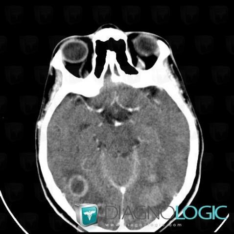
Here is the specific information in the key image above:
- Diagnosis Metastasis, Location(s) Cerebral hemispheres, with gamuts Intracerebral lesion with ring enhancement, Intracerebral lesion with moderate enhancement, Multifocal intracranial lesions
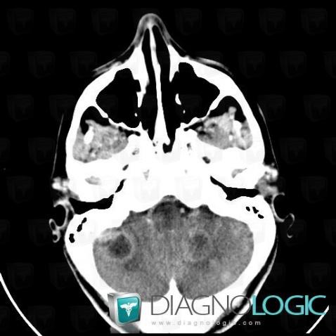
Here is the specific information in the key image above:
- Diagnosis Metastasis, Location(s) Posterior fossa, with gamuts Infratentorial cystic mass mass with a mural nodule, Infratentorial lesion with ring enhancement, Infratentorial lesion with moderate enhancement, Multifocal infratentorial lesionsCerebellar hemisphere, with gamuts Cerebellar lesion
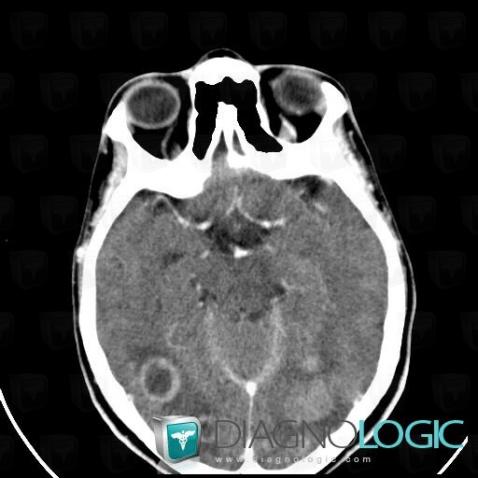
Here is the specific information in the key image above:
- Diagnosis Metastasis, Location(s) Cerebral hemispheres, with gamuts Intracerebral lesion with ring enhancement, Intracerebral lesion with moderate enhancement, Multifocal intracranial lesions
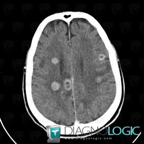
Here is the specific information in the key image above:
- Diagnosis Metastasis, Location(s) Cortico subcortical region, with gamuts Subcortical lesionCerebral hemispheres, with gamuts Intracerebral lesion with intense enhancement
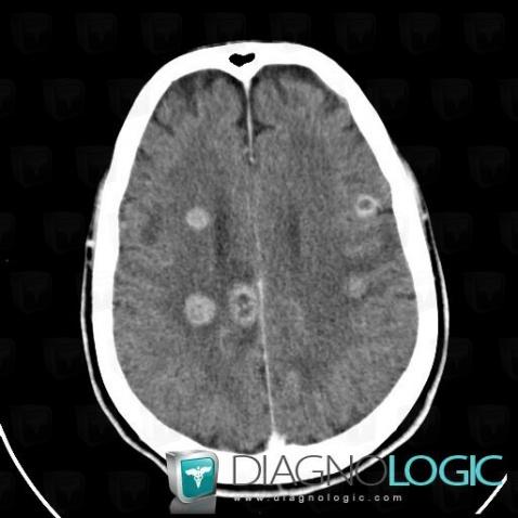
Here is the specific information in the key image above:
- Diagnosis Metastasis, Location(s) Cortico subcortical region, with gamuts Subcortical lesionCerebral hemispheres, with gamuts Intracerebral lesion with intense enhancement