The images below illustrate this case for diagnoses Parathroid adenoma (link to Parathyroid lesion), Hyperparathyroidism, Brown tumor of hyperparathyroidism, for the modalities (CT ,MRI ,X rays)
Gamuts for this case : :
- Lytic lesion in a vertebra
- Osteolytic lesion of ribs
- Solid cervical mass
- Anterior mediastinal mass
- Superior mediastinal mass
- Without gamut
- Posterior arch lesion
- Generalised osteosclerosis
- Expansive lesion
- Mulltiple osteolysis
- Well-defined osteolysis
- Meta-diaphyseal osteolysis
- Bilateral symmetrical sacroiliac disease
- T2 WI Hypointense bone lesion
- Markedly hyperintense T2 WI bone lesion
- Diaphyseal osteolysis
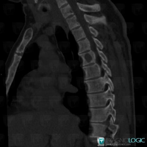
Here is the specific information in the key image above:
- Diagnosis Brown tumor of hyperparathyroidism, Location(s) Vertebral body / Disk, with gamuts Lytic lesion in a vertebra
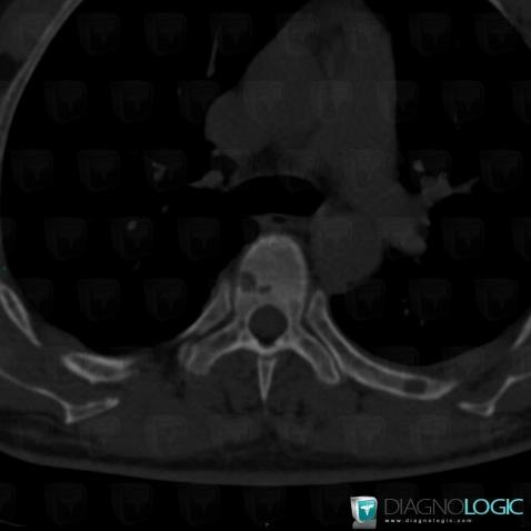
Here is the specific information in the key image above:
- Diagnosis Brown tumor of hyperparathyroidism, Location(s) Ribs, with gamuts Osteolytic lesion of ribs
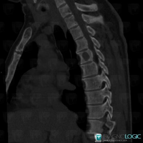
Here is the specific information in the key image above:
- Diagnosis Brown tumor of hyperparathyroidism, Location(s) Vertebral body / Disk, with gamuts Lytic lesion in a vertebra
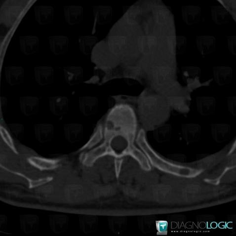
Here is the specific information in the key image above:
- Diagnosis Brown tumor of hyperparathyroidism, Location(s) Ribs, with gamuts Osteolytic lesion of ribs
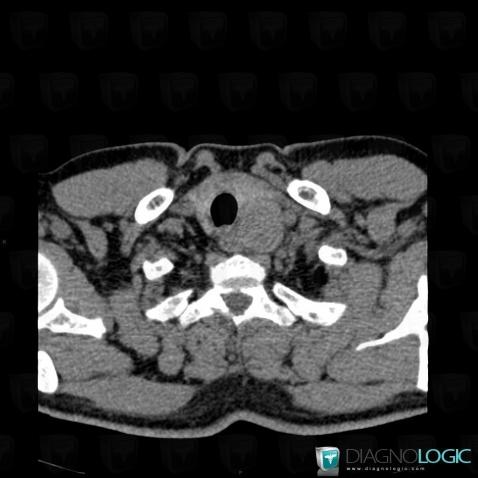
Here is the specific information in the key image above:
- Diagnosis Parathroid adenoma (link to Parathyroid lesion), Location(s) Deep neck spaces, with gamuts Solid cervical mass
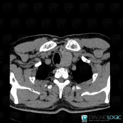
Here is the specific information in the key image above:
- Diagnosis Parathroid adenoma (link to Parathyroid lesion), Location(s) Mediastinum, with gamuts Anterior mediastinal mass, Superior mediastinal mass
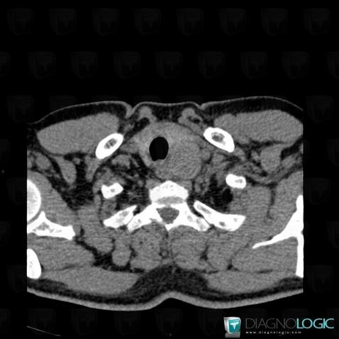
Here is the specific information in the key image above:
- Diagnosis Parathroid adenoma (link to Parathyroid lesion), Location(s) Deep neck spaces, with gamuts Solid cervical mass
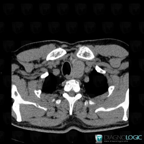
Here is the specific information in the key image above:
- Diagnosis Parathroid adenoma (link to Parathyroid lesion), Location(s) Mediastinum, with gamuts Anterior mediastinal mass, Superior mediastinal mass
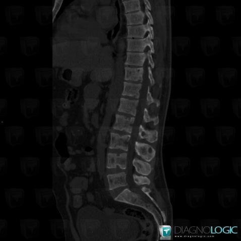
Here is the specific information in the key image above:
- Diagnosis Hyperparathyroidism, Location(s) Vertebral body / Disk, with gamuts
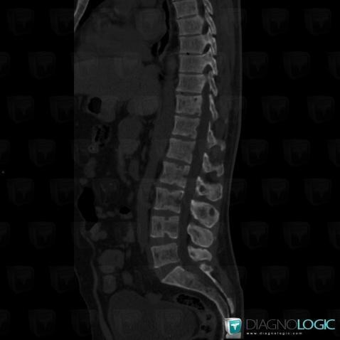
Here is the specific information in the key image above:
- Diagnosis Hyperparathyroidism, Location(s) Vertebral body / Disk, with gamuts
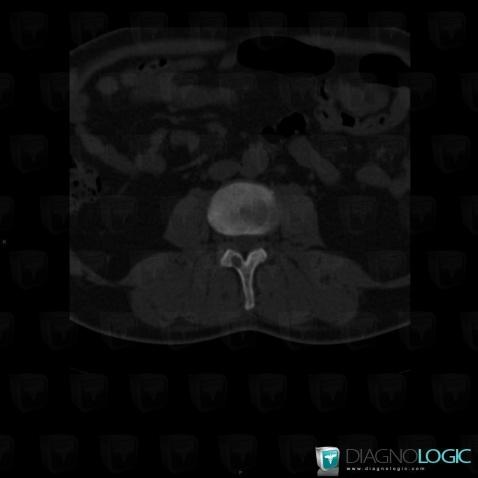
Here is the specific information in the key image above:
- Diagnosis Brown tumor of hyperparathyroidism, Location(s) Vertebral body / Disk, with gamuts Posterior arch lesion
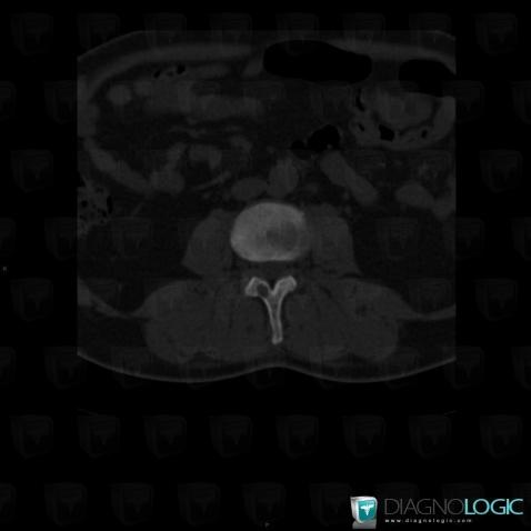
Here is the specific information in the key image above:
- Diagnosis Brown tumor of hyperparathyroidism, Location(s) Vertebral body / Disk, with gamuts Posterior arch lesion
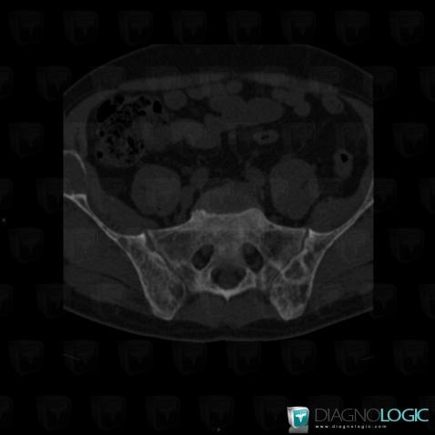
Here is the specific information in the key image above:
- Diagnosis Hyperparathyroidism, Location(s) Ilium, with gamuts Generalised osteosclerosis
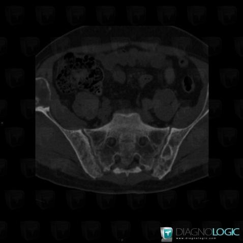
Here is the specific information in the key image above:
- Diagnosis Brown tumor of hyperparathyroidism, Location(s) Ilium, with gamuts Expansive lesion
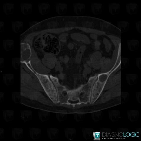
Here is the specific information in the key image above:
- Diagnosis Brown tumor of hyperparathyroidism, Location(s) Ilium, with gamuts Expansive lesion, Mulltiple osteolysis
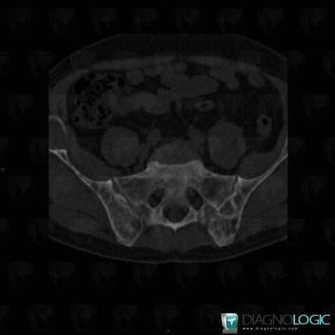
Here is the specific information in the key image above:
- Diagnosis Hyperparathyroidism, Location(s) Ilium, with gamuts Generalised osteosclerosis
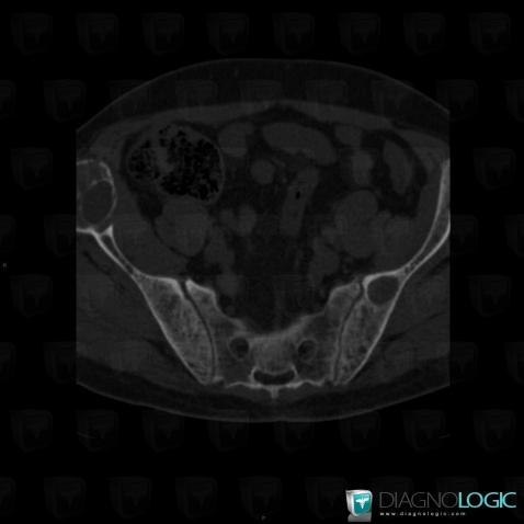
Here is the specific information in the key image above:
- Diagnosis Brown tumor of hyperparathyroidism, Location(s) Ilium, with gamuts Expansive lesion, Mulltiple osteolysis
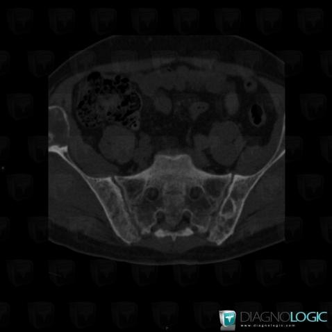
Here is the specific information in the key image above:
- Diagnosis Brown tumor of hyperparathyroidism, Location(s) Ilium, with gamuts Expansive lesion
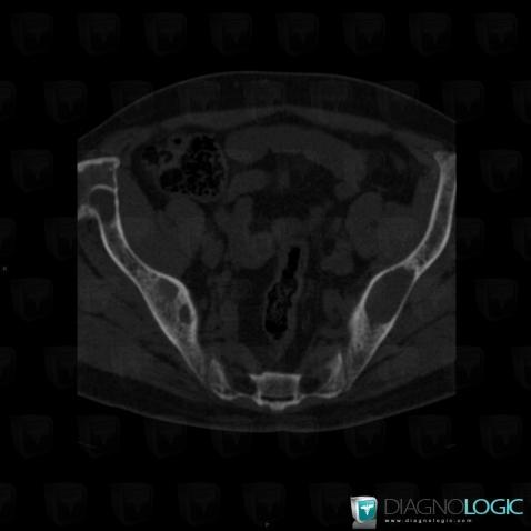
Here is the specific information in the key image above:
- Diagnosis Brown tumor of hyperparathyroidism, Location(s) Ilium, with gamuts Well-defined osteolysis
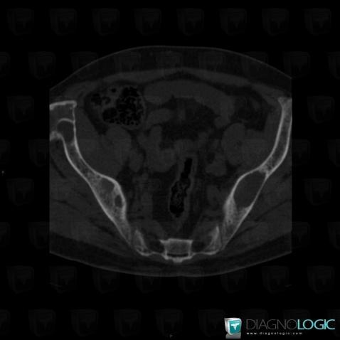
Here is the specific information in the key image above:
- Diagnosis Brown tumor of hyperparathyroidism, Location(s) Ilium, with gamuts Well-defined osteolysis
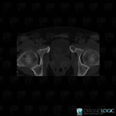
Here is the specific information in the key image above:
- Diagnosis Brown tumor of hyperparathyroidism, Location(s) Femur - Proximal part, with gamuts Mulltiple osteolysis, Meta-diaphyseal osteolysis, Well-defined osteolysis
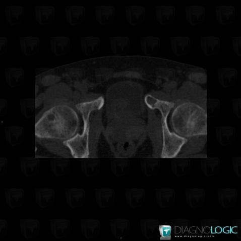
Here is the specific information in the key image above:
- Diagnosis Brown tumor of hyperparathyroidism, Location(s) Femur - Proximal part, with gamuts Mulltiple osteolysis, Meta-diaphyseal osteolysis, Well-defined osteolysis
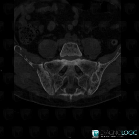
Here is the specific information in the key image above:
- Diagnosis Hyperparathyroidism, Location(s) Sacro iliac joint, with gamuts Bilateral symmetrical sacroiliac disease
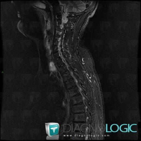
Here is the specific information in the key image above:
- Diagnosis Brown tumor of hyperparathyroidism, Location(s) Vertebral body / Disk, with gamuts T2 WI Hypointense bone lesion
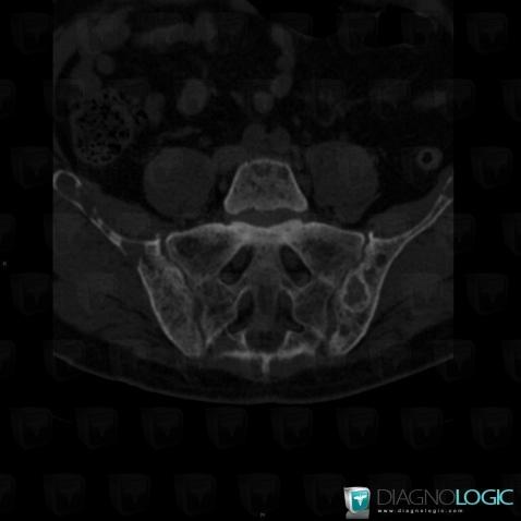
Here is the specific information in the key image above:
- Diagnosis Hyperparathyroidism, Location(s) Sacro iliac joint, with gamuts Bilateral symmetrical sacroiliac disease
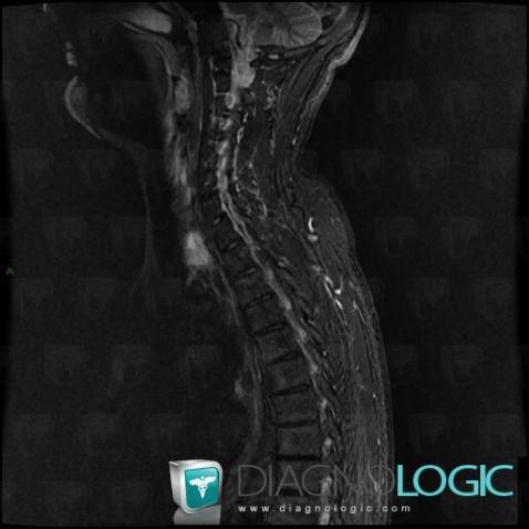
Here is the specific information in the key image above:
- Diagnosis Brown tumor of hyperparathyroidism, Location(s) Vertebral body / Disk, with gamuts T2 WI Hypointense bone lesion
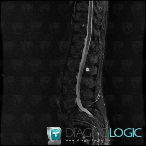
Here is the specific information in the key image above:
- Diagnosis Brown tumor of hyperparathyroidism, Location(s) Vertebral body / Disk, with gamuts Markedly hyperintense T2 WI bone lesion
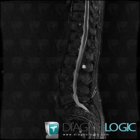
Here is the specific information in the key image above:
- Diagnosis Brown tumor of hyperparathyroidism, Location(s) Vertebral body / Disk, with gamuts Markedly hyperintense T2 WI bone lesion
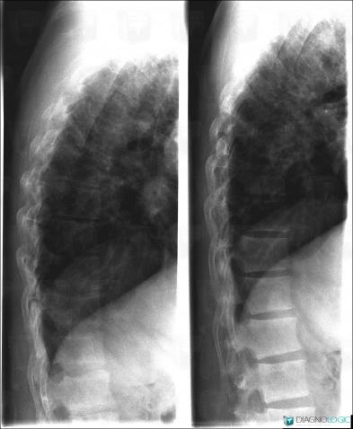
Here is the specific information in the key image above:
- Diagnosis Brown tumor of hyperparathyroidism, Location(s) Vertebral body / Disk, with gamuts Lytic lesion in a vertebra
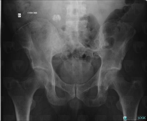
Here is the specific information in the key image above:
- Diagnosis Hyperparathyroidism, Location(s) Ilium, with gamuts Generalised osteosclerosis
- Diagnosis Brown tumor of hyperparathyroidism, Location(s) Ilium, with gamuts Mulltiple osteolysis, Well-defined osteolysis, Expansive lesion
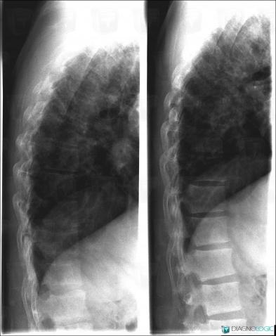
Here is the specific information in the key image above:
- Diagnosis Brown tumor of hyperparathyroidism, Location(s) Vertebral body / Disk, with gamuts Lytic lesion in a vertebra
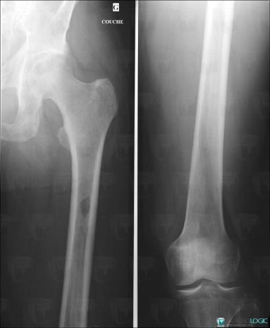
Here is the specific information in the key image above:
- Diagnosis Hyperparathyroidism, Location(s) Femur - Mid part, with gamuts Generalised osteosclerosisFemur - Distal part, with gamuts Generalised osteosclerosisFemur - Proximal part, with gamuts Generalised osteosclerosis
- Diagnosis Brown tumor of hyperparathyroidism, Location(s) Fibula - Mid part, with gamuts Diaphyseal osteolysis, Well-defined osteolysis, Mulltiple osteolysis
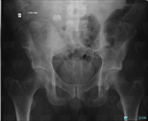
Here is the specific information in the key image above:
- Diagnosis Hyperparathyroidism, Location(s) Ilium, with gamuts Generalised osteosclerosis
- Diagnosis Brown tumor of hyperparathyroidism, Location(s) Ilium, with gamuts Mulltiple osteolysis, Well-defined osteolysis, Expansive lesion
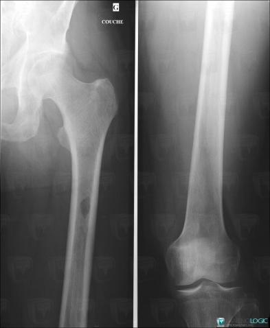
Here is the specific information in the key image above:
- Diagnosis Hyperparathyroidism, Location(s) Femur - Mid part, with gamuts Generalised osteosclerosisFemur - Distal part, with gamuts Generalised osteosclerosisFemur - Proximal part, with gamuts Generalised osteosclerosis
- Diagnosis Brown tumor of hyperparathyroidism, Location(s) Fibula - Mid part, with gamuts Diaphyseal osteolysis, Well-defined osteolysis, Mulltiple osteolysis