The images below illustrate this case for diagnoses Multiple myeloma, for the modalities (CT ,X rays ,US)
Gamuts for this case : :
- Low density retroperitoneal mass
- Solid pelvic mass
- Large pelvic mass
- Presacral mass
- Without gamut
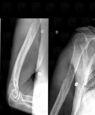
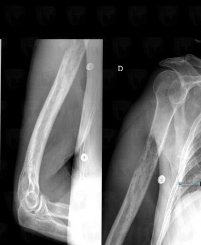
- Motheaten or permeative lesion
- Diaphyseal osteolysis
- Ill-defined osteolysis
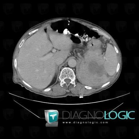
Here is the specific information in the key image above:
- Diagnosis Multiple myeloma, Location(s) Retroperitoneum, with gamuts Low density retroperitoneal mass
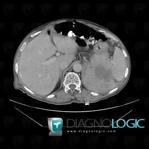
Here is the specific information in the key image above:
- Diagnosis Multiple myeloma, Location(s) Retroperitoneum, with gamuts Low density retroperitoneal mass
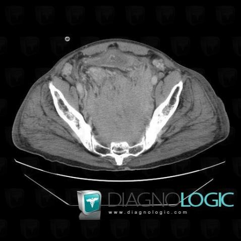
Here is the specific information in the key image above:
- Diagnosis Multiple myeloma, Location(s) Pelvis / Perineum, with gamuts Solid pelvic mass, Large pelvic mass, Presacral mass
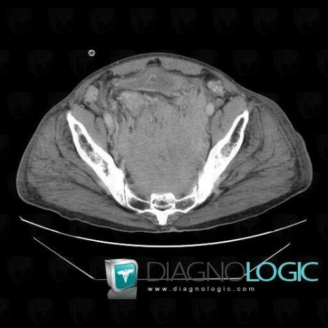
Here is the specific information in the key image above:
- Diagnosis Multiple myeloma, Location(s) Pelvis / Perineum, with gamuts Solid pelvic mass, Large pelvic mass, Presacral mass
Here is the specific information in the key image above:
Here is the specific information in the key image above:
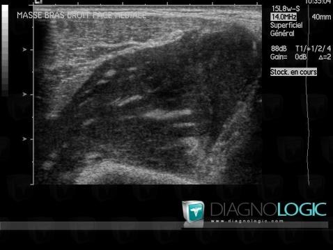
- Diagnosis Multiple myeloma, Location(s) Other soft tissues/nerves - Arm, with gamuts
Here is the specific information in the key image above:
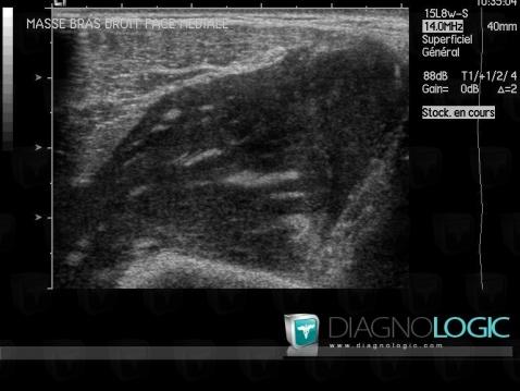
- Diagnosis Multiple myeloma, Location(s) Other soft tissues/nerves - Arm, with gamuts
Here is the specific information in the key image above:
- Diagnosis Multiple myeloma, Location(s) Humerus - Mid part, with gamuts Motheaten or permeative lesion, Diaphyseal osteolysis, Ill-defined osteolysis