The images below illustrate this case for diagnoses Metastasis, Lymphangitic carcinomatosis, Breast carcinoma, for the modalities (CT)
Gamuts for this case : :
- Chest wall lesion
- Low density in portal phase hepatic lesion
- Solid hepatic lesion
- Perilymphatic micronodules
- Irregular peribronchovacular / interlobular thickening
- Peribronchovacular / interlobular thickening
- Focal area of sclerosis in a vertebra
- Multiple osteosclerotic bone lesions
- Sacral lesion
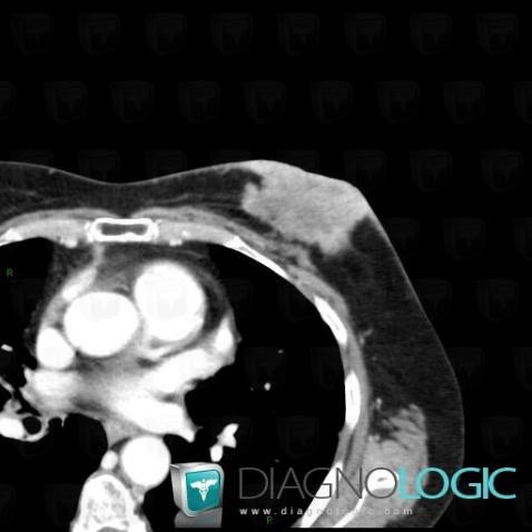
Here is the specific information in the key image above:
- Diagnosis Breast carcinoma, Location(s) Chest wall, with gamuts Chest wall lesion
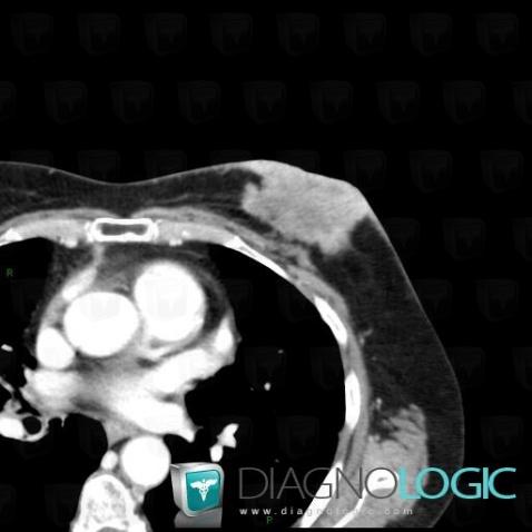
Here is the specific information in the key image above:
- Diagnosis Breast carcinoma, Location(s) Chest wall, with gamuts Chest wall lesion
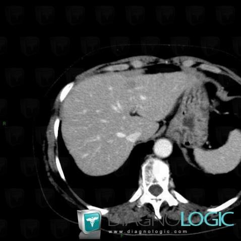
Here is the specific information in the key image above:
- Diagnosis Metastasis, Location(s) Liver, with gamuts Low density in portal phase hepatic lesion, Solid hepatic lesion
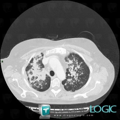
Here is the specific information in the key image above:
- Diagnosis Lymphangitic carcinomatosis, Location(s) Pulmonary parenchyma, with gamuts Perilymphatic micronodules, Irregular peribronchovacular / interlobular thickening, Peribronchovacular / interlobular thickening
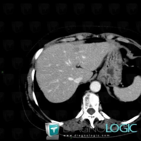
Here is the specific information in the key image above:
- Diagnosis Metastasis, Location(s) Liver, with gamuts Low density in portal phase hepatic lesion, Solid hepatic lesion
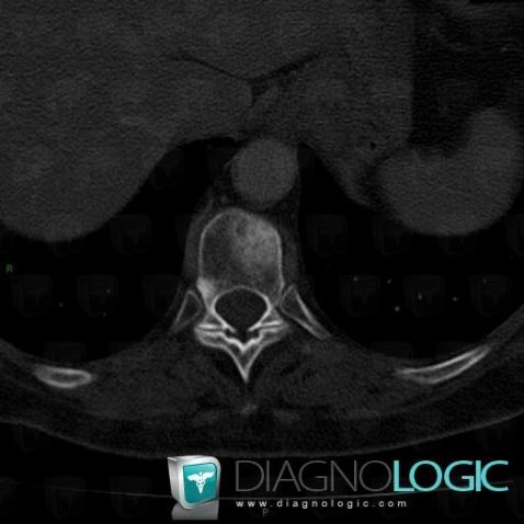
Here is the specific information in the key image above:
- Diagnosis Metastasis, Location(s) Vertebral body / Disk, with gamuts Focal area of sclerosis in a vertebra
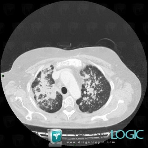
Here is the specific information in the key image above:
- Diagnosis Lymphangitic carcinomatosis, Location(s) Pulmonary parenchyma, with gamuts Perilymphatic micronodules, Irregular peribronchovacular / interlobular thickening, Peribronchovacular / interlobular thickening
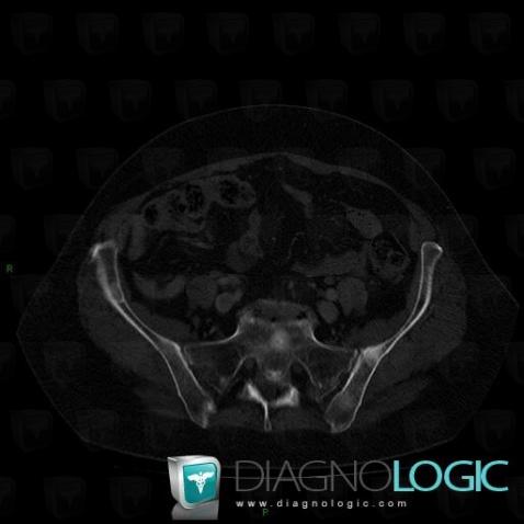
Here is the specific information in the key image above:
- Diagnosis Metastasis, Location(s) Ilium, with gamuts Multiple osteosclerotic bone lesionsSacrum / Coccyx, with gamuts Sacral lesion
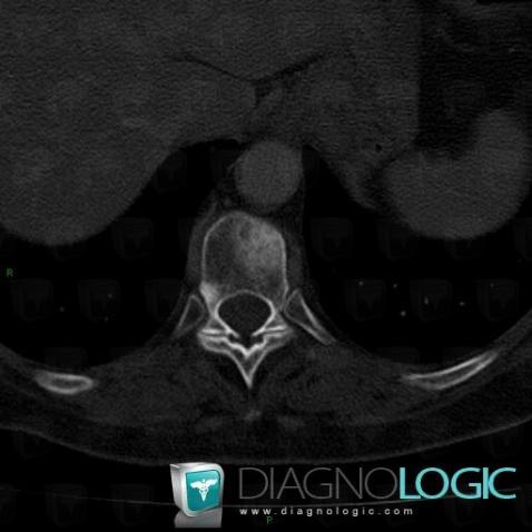
Here is the specific information in the key image above:
- Diagnosis Metastasis, Location(s) Vertebral body / Disk, with gamuts Focal area of sclerosis in a vertebra
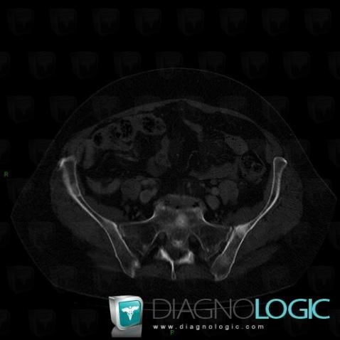
Here is the specific information in the key image above:
- Diagnosis Metastasis, Location(s) Ilium, with gamuts Multiple osteosclerotic bone lesionsSacrum / Coccyx, with gamuts Sacral lesion