The images below illustrate this case for diagnoses Meningioma, for the modalities (CT)
Gamuts for this case : :
- Optic nerve lesions
- Intracerebral lesion with intense enhancement
- Focal meningeal enhancement
- Supratentorial extra axial lesion
- Cavernous sinus lesion
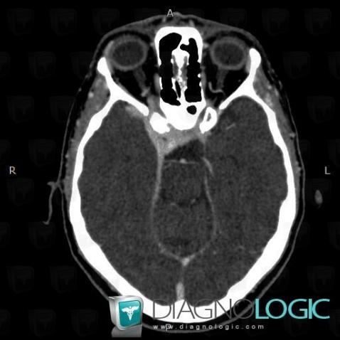
Here is the specific information in the key image above:
- Diagnosis Meningioma, Location(s) Optic nerve, with gamuts Optic nerve lesions
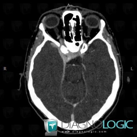
Here is the specific information in the key image above:
- Diagnosis Meningioma, Location(s) Optic nerve, with gamuts Optic nerve lesions
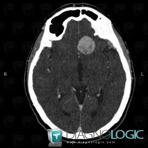
Here is the specific information in the key image above:
- Diagnosis Meningioma, Location(s) Cerebral hemispheres, with gamuts Intracerebral lesion with intense enhancement
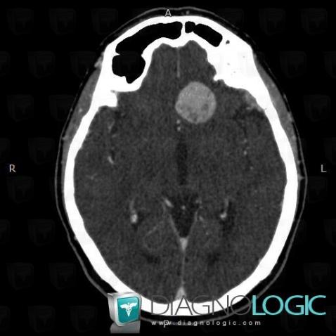
Here is the specific information in the key image above:
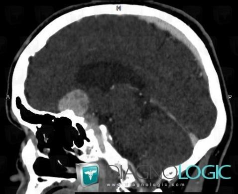
Here is the specific information in the key image above:
- Diagnosis Meningioma, Location(s) Supratentorial peri cerebral spaces, with gamuts Focal meningeal enhancement, Supratentorial extra axial lesion
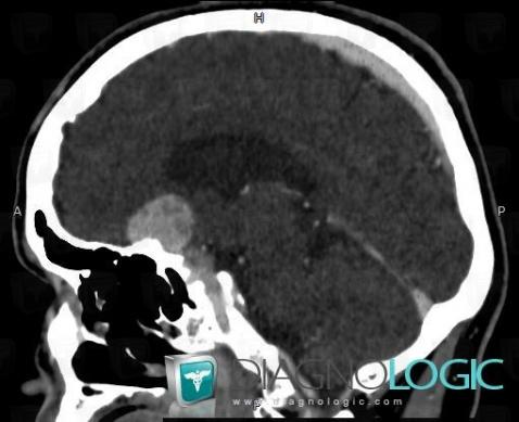
Here is the specific information in the key image above:
- Diagnosis Meningioma, Location(s) Supratentorial peri cerebral spaces, with gamuts Focal meningeal enhancement, Supratentorial extra axial lesion
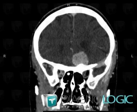
Here is the specific information in the key image above:
- Diagnosis Meningioma, Location(s) Eye, with gamuts Optic nerve lesions
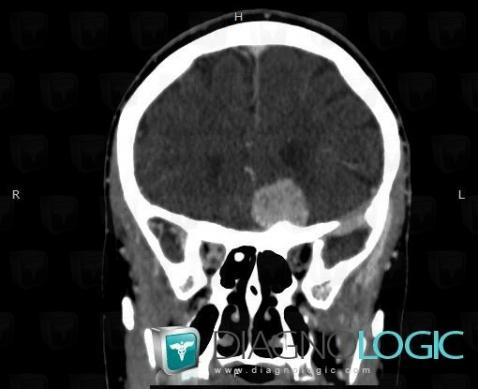
Here is the specific information in the key image above:
- Diagnosis Meningioma, Location(s) Eye, with gamuts Optic nerve lesions
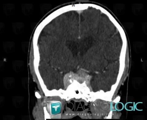
Here is the specific information in the key image above:
- Diagnosis Meningioma, Location(s) Pituitary gland and parasellar region, with gamuts Cavernous sinus lesion
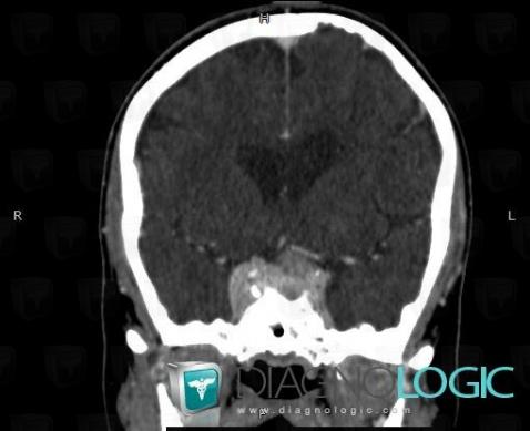
Here is the specific information in the key image above:
- Diagnosis Meningioma, Location(s) Pituitary gland and parasellar region, with gamuts Cavernous sinus lesion