The images below illustrate this case for diagnoses Meningioma, for the modalities (MRI)
Gamuts for this case : :
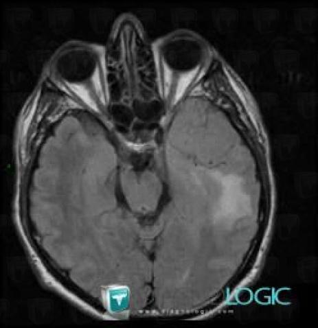
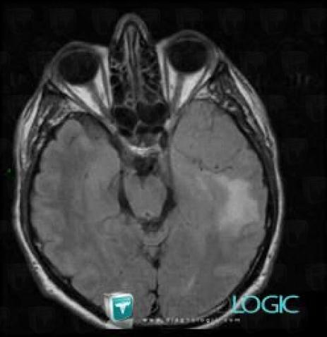
- Intracerebral T2W or FLAIR isointense lesion
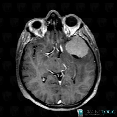
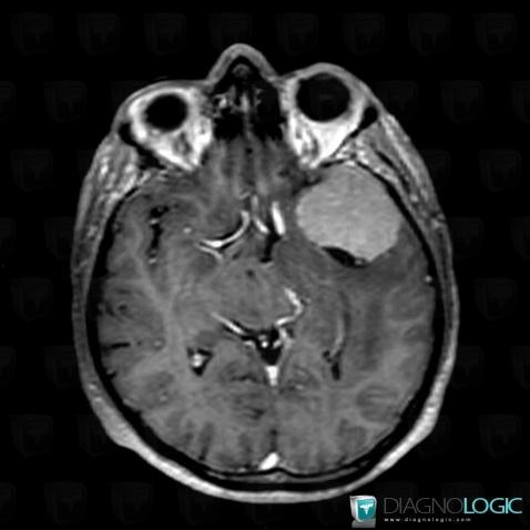
- Intracerebral lesion with intense enhancement
- Focal meningeal enhancement
- Supratentorial extra axial lesion

Here is the specific information in the key image above:
- Diagnosis Meningioma, Location(s) Cerebral hemispheres, with gamuts Intracerebral T2W or FLAIR isointense lesion

Here is the specific information in the key image above:
- Diagnosis Meningioma, Location(s) Cerebral hemispheres, with gamuts Intracerebral T2W or FLAIR isointense lesion

Here is the specific information in the key image above:
- Diagnosis Meningioma, Location(s) Cerebral hemispheres, with gamuts Intracerebral lesion with intense enhancementSupratentorial peri cerebral spaces, with gamuts Focal meningeal enhancement, Supratentorial extra axial lesion

Here is the specific information in the key image above:
- Diagnosis Meningioma, Location(s) Cerebral hemispheres, with gamuts Intracerebral lesion with intense enhancementSupratentorial peri cerebral spaces, with gamuts Focal meningeal enhancement, Supratentorial extra axial lesion