The images below illustrate this case for diagnoses Lymphoma, for the modalities (MRI ,CT)
Gamuts for this case : :
- Low attenuation mediastinal mass
- Middle mediastinal mass
- Hilar lymph nodes enlargement
- Without gamut
- Lesion in the perinephric space
- Paraspinal soft tissue mass
- Small bowel mass or filling defect
- Lesion in the psoas space
- Focal thicknening of small bowel wall
- Extradural lesion
- Focal meningeal enhancement
- Supratentorial extra axial lesion
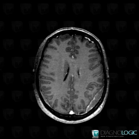
Here is the specific information in the key image above:
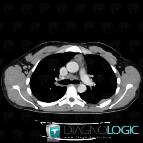
Here is the specific information in the key image above:
- Diagnosis Lymphoma, Location(s) Mediastinum, with gamuts Low attenuation mediastinal mass, Middle mediastinal mass
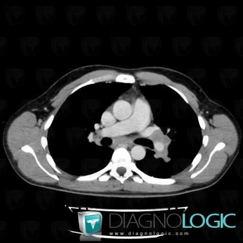
Here is the specific information in the key image above:
- Diagnosis Lymphoma, Location(s) Mediastinum, with gamuts Hilar lymph nodes enlargement
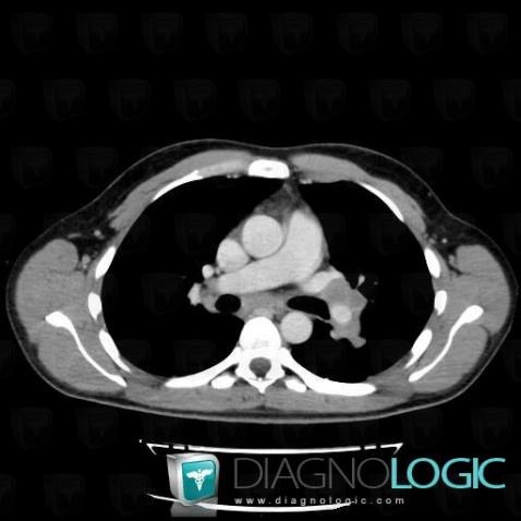
Here is the specific information in the key image above:
- Diagnosis Lymphoma, Location(s) Mediastinum, with gamuts Hilar lymph nodes enlargement
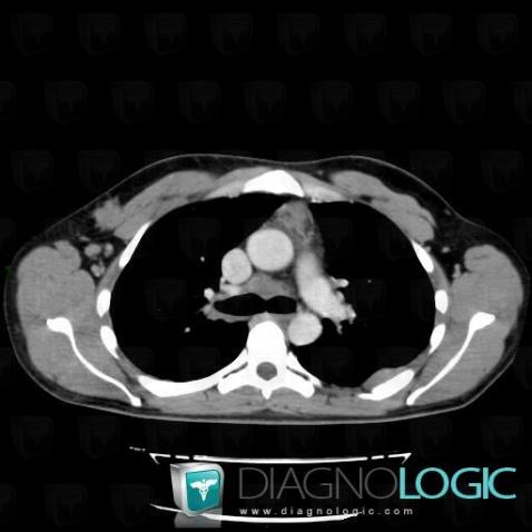
Here is the specific information in the key image above:
- Diagnosis Lymphoma, Location(s) Mediastinum, with gamuts Low attenuation mediastinal mass, Middle mediastinal mass
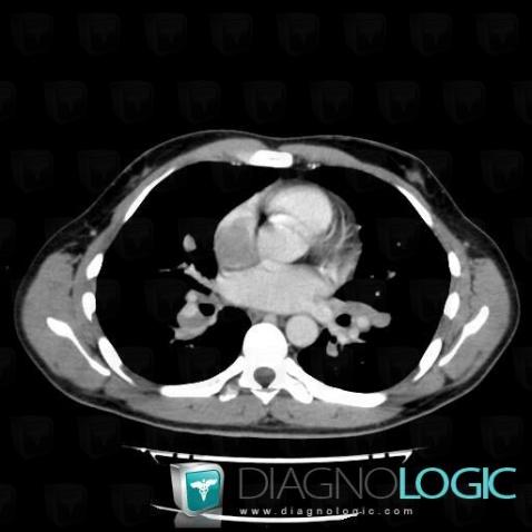
Here is the specific information in the key image above:
- Diagnosis Lymphoma, Location(s) Cardiac cavities / Pericardium, with gamuts
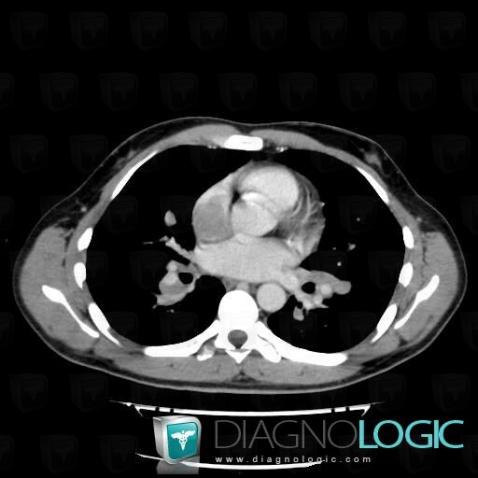
Here is the specific information in the key image above:
- Diagnosis Lymphoma, Location(s) Cardiac cavities / Pericardium, with gamuts
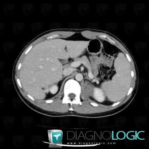
Here is the specific information in the key image above:
- Diagnosis Lymphoma, Location(s) Retroperitoneum, with gamuts Lesion in the perinephric space
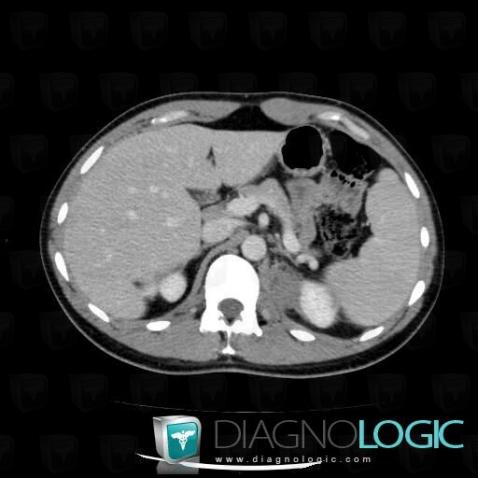
Here is the specific information in the key image above:
- Diagnosis Lymphoma, Location(s) Retroperitoneum, with gamuts Lesion in the perinephric space
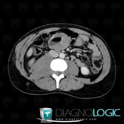
Here is the specific information in the key image above:
- Diagnosis Lymphoma, Location(s) Paraspinal, with gamuts Paraspinal soft tissue massSmall bowel, with gamuts Small bowel mass or filling defect, Focal thicknening of small bowel wallRetroperitoneum, with gamuts Lesion in the psoas space
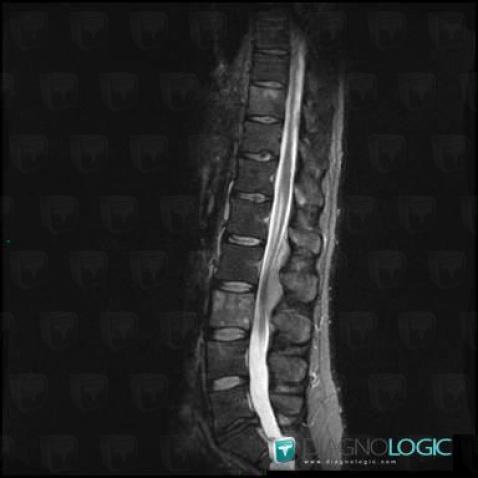
Here is the specific information in the key image above:
- Diagnosis Lymphoma, Location(s) Spinal canal / Cord, with gamuts Extradural lesionVertebral body / Disk, with gamuts
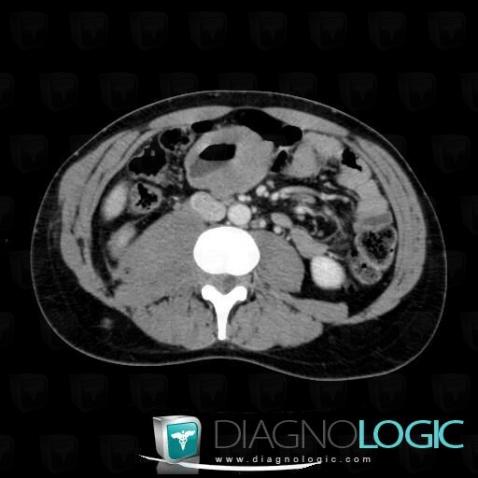
Here is the specific information in the key image above:
- Diagnosis Lymphoma, Location(s) Paraspinal, with gamuts Paraspinal soft tissue massSmall bowel, with gamuts Small bowel mass or filling defect, Focal thicknening of small bowel wallRetroperitoneum, with gamuts Lesion in the psoas space
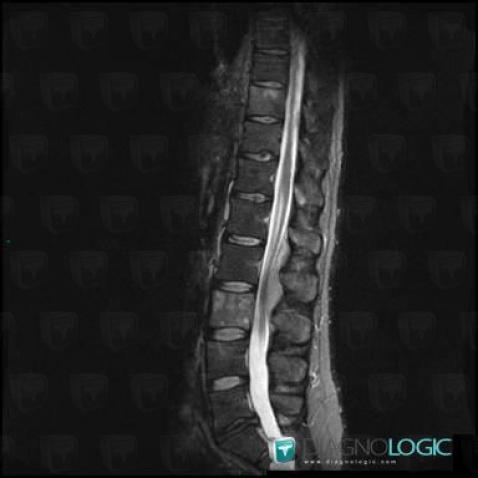
Here is the specific information in the key image above:
- Diagnosis Lymphoma, Location(s) Spinal canal / Cord, with gamuts Extradural lesionVertebral body / Disk, with gamuts
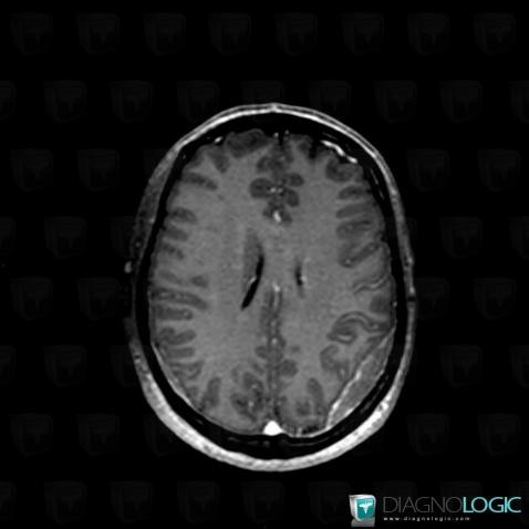
Here is the specific information in the key image above:
- Diagnosis Lymphoma, Location(s) Supratentorial peri cerebral spaces, with gamuts Focal meningeal enhancement, Supratentorial extra axial lesion
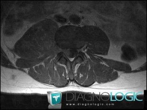
Here is the specific information in the key image above:
- Diagnosis Lymphoma, Location(s) Paraspinal, with gamuts Paraspinal soft tissue massRetroperitoneum, with gamuts Lesion in the psoas space
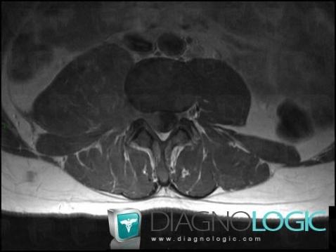
Here is the specific information in the key image above:
- Diagnosis Lymphoma, Location(s) Paraspinal, with gamuts Paraspinal soft tissue massRetroperitoneum, with gamuts Lesion in the psoas space