The images below illustrate this case for diagnoses Lymphoma, Peritoneal carcinomatosis, for the modalities (CT)
Gamuts for this case : :
- Cardiophrenic angle mass
- Peritoneal disease
- Marked thicknening of small bowel wall
- Terminal ileum lesion
- Small bowel mass or filling defect
- Small bowel fold thickening
- Focal thicknening of small bowel wall
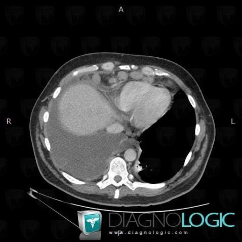
Here is the specific information in the key image above:
- Diagnosis Peritoneal carcinomatosis, Location(s) Mediastinum, with gamuts Cardiophrenic angle mass
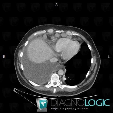
Here is the specific information in the key image above:
- Diagnosis Peritoneal carcinomatosis, Location(s) Mediastinum, with gamuts Cardiophrenic angle mass
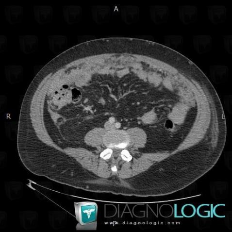
Here is the specific information in the key image above:
- Diagnosis Peritoneal carcinomatosis, Location(s) Mesentery / Peritoneum, with gamuts Peritoneal disease
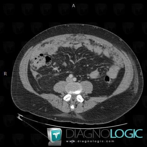
Here is the specific information in the key image above:
- Diagnosis Peritoneal carcinomatosis, Location(s) Mesentery / Peritoneum, with gamuts Peritoneal disease
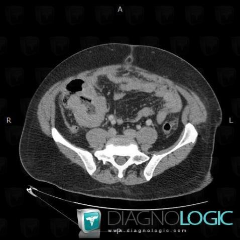
Here is the specific information in the key image above:
- Diagnosis Lymphoma, Location(s) Small bowel, with gamuts Marked thicknening of small bowel wall, Terminal ileum lesion, Small bowel mass or filling defect, Small bowel fold thickening, Focal thicknening of small bowel wall
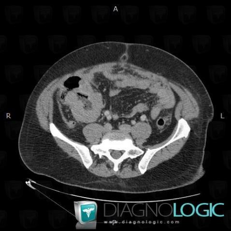
Here is the specific information in the key image above:
- Diagnosis Lymphoma, Location(s) Small bowel, with gamuts Marked thicknening of small bowel wall, Terminal ileum lesion, Small bowel mass or filling defect, Small bowel fold thickening, Focal thicknening of small bowel wall