The images below illustrate this case for diagnoses Lymphoma, for the modalities (CT ,MRI)
Gamuts for this case : :
- Hyperdense intracerebral lesion on noncontrast CT
- Isodense intracerebral lesion on noncontrast CT
- Periventricular hypodensity
- Multifocal intracranial lesions
- Without gamut
- Intracerebral lesion with intense enhancement
- Intracerebral mass
- Midline mass
- Gyriform anomaly
- Intracerebral lesion with ring enhancement
- Subcortical lesion
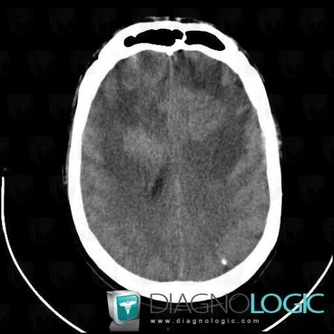
Here is the specific information in the key image above:
- Diagnosis Lymphoma, Location(s) Cerebral hemispheres, with gamuts Hyperdense intracerebral lesion on noncontrast CT
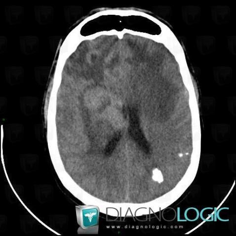
Here is the specific information in the key image above:
- Diagnosis Lymphoma, Location(s) Cerebral hemispheres, with gamuts Isodense intracerebral lesion on noncontrast CTVentricles / Periventricular region, with gamuts Periventricular hypodensity
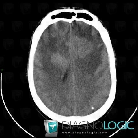
Here is the specific information in the key image above:
- Diagnosis Lymphoma, Location(s) Cerebral hemispheres, with gamuts Hyperdense intracerebral lesion on noncontrast CT
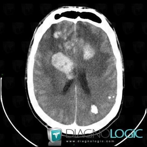
Here is the specific information in the key image above:
- Diagnosis Lymphoma, Location(s) Cerebral hemispheres, with gamuts Multifocal intracranial lesions, Intracerebral lesion with intense enhancement, Intracerebral massBasal ganglia and capsule, with gamuts
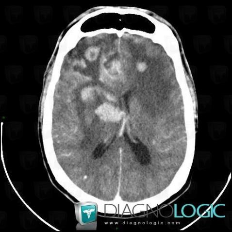
Here is the specific information in the key image above:
- Diagnosis Lymphoma, Location(s) Cerebral falx / Midline, with gamuts Midline massCortico subcortical region, with gamuts Gyriform anomaly, Subcortical lesionCerebral hemispheres, with gamuts Intracerebral lesion with ring enhancement
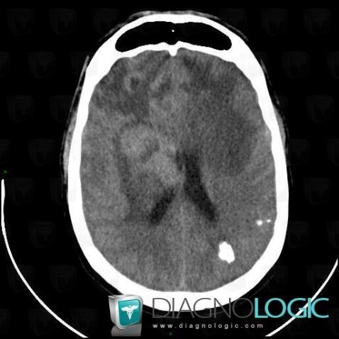
Here is the specific information in the key image above:
- Diagnosis Lymphoma, Location(s) Cerebral hemispheres, with gamuts Isodense intracerebral lesion on noncontrast CTVentricles / Periventricular region, with gamuts Periventricular hypodensity
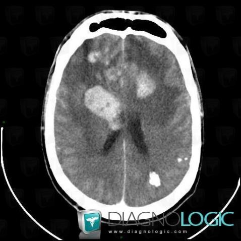
Here is the specific information in the key image above:
- Diagnosis Lymphoma, Location(s) Cerebral hemispheres, with gamuts Multifocal intracranial lesions, Intracerebral lesion with intense enhancement, Intracerebral massBasal ganglia and capsule, with gamuts
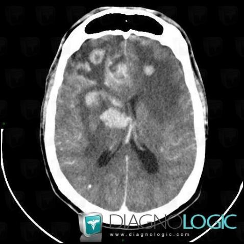
Here is the specific information in the key image above:
- Diagnosis Lymphoma, Location(s) Cerebral falx / Midline, with gamuts Midline massCortico subcortical region, with gamuts Gyriform anomaly, Subcortical lesionCerebral hemispheres, with gamuts Intracerebral lesion with ring enhancement