The images below illustrate this case for diagnoses Lymphocele, for the modalities (MRI ,CT)
Gamuts for this case : :
- Cystic retroperitoneal mass
- Lesion in the psoas space
- T2 WI hyperintense adnexal mass
- Cystic adnexal mass
- Low density retroperitoneal mass
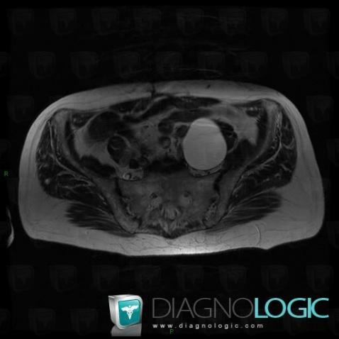
Here is the specific information in the key image above:
- Diagnosis Lymphocele, Location(s) Retroperitoneum, with gamuts Cystic retroperitoneal mass, Lesion in the psoas space
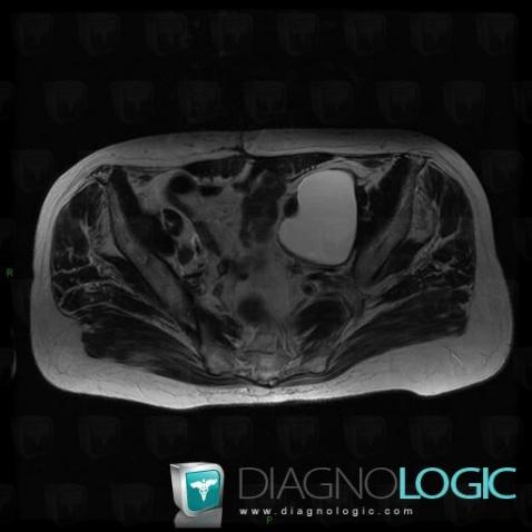
Here is the specific information in the key image above:
- Diagnosis Lymphocele, Location(s) Adnexa / Ovary fallopian tube, with gamuts T2 WI hyperintense adnexal mass, Cystic adnexal mass
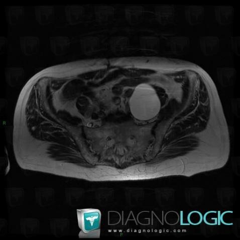
Here is the specific information in the key image above:
- Diagnosis Lymphocele, Location(s) Retroperitoneum, with gamuts Cystic retroperitoneal mass, Lesion in the psoas space
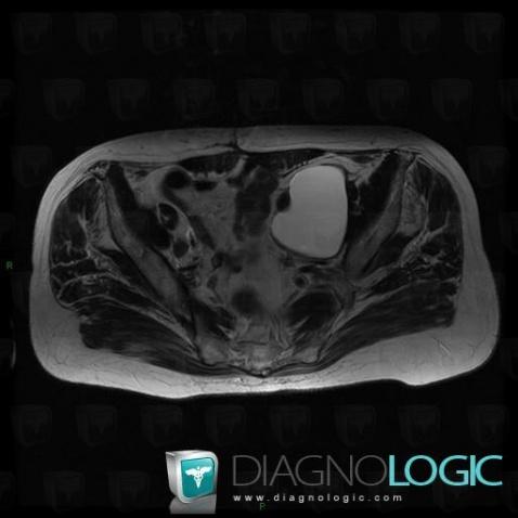
Here is the specific information in the key image above:
- Diagnosis Lymphocele, Location(s) Adnexa / Ovary fallopian tube, with gamuts T2 WI hyperintense adnexal mass, Cystic adnexal mass
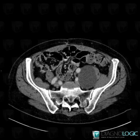
Here is the specific information in the key image above:
- Diagnosis Lymphocele, Location(s) Retroperitoneum, with gamuts Lesion in the psoas space, Cystic retroperitoneal mass, Low density retroperitoneal mass
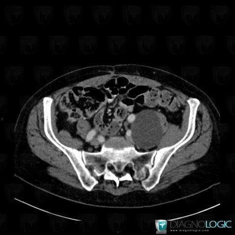
Here is the specific information in the key image above:
- Diagnosis Lymphocele, Location(s) Retroperitoneum, with gamuts Lesion in the psoas space, Cystic retroperitoneal mass, Low density retroperitoneal mass