The images below illustrate this case for diagnoses Low grade astrocytoma, for the modalities (CT ,MRI)
Gamuts for this case : :
- Basal ganglia calcifications
- Hypodense intracerebral lesion on noncontrast CT
- Basal ganglia hypodense lesion
- Periventricular hypodensity
- Intracerebral mass
- Intraventricular mass
- Intracerebral T2W or FLAIR hyperintense lesion
- Basal ganglia T2W or FLAIR hyperintense lesion
- Subcortical lesion
- Cortical lesion
- Periventricular anomaly seen in MRI
- Ventricular wall nodules
- Corpus callosum lesion
- DWI hyperintense lesion
- Intracerebral lesion with moderate enhancement
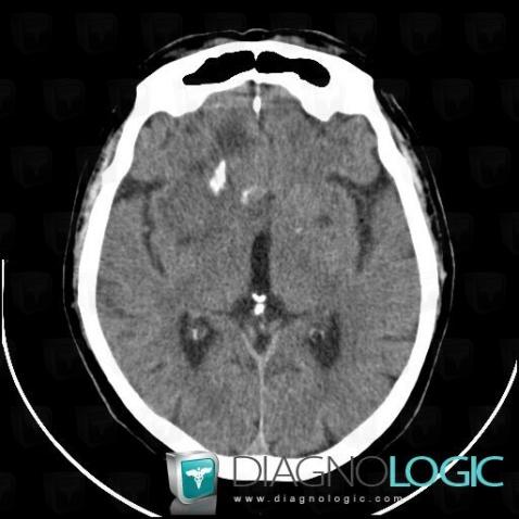
Here is the specific information in the key image above:
- Diagnosis Low grade astrocytoma, Location(s) Basal ganglia and capsule, with gamuts Basal ganglia calcifications, Basal ganglia hypodense lesionCerebral hemispheres, with gamuts Hypodense intracerebral lesion on noncontrast CT, Intracerebral massVentricles / Periventricular region, with gamuts Periventricular hypodensity
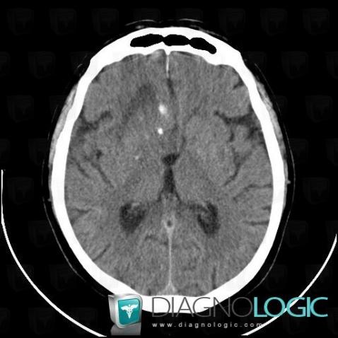
Here is the specific information in the key image above:
- Diagnosis Low grade astrocytoma, Location(s) Ventricles / Periventricular region, with gamuts Intraventricular mass
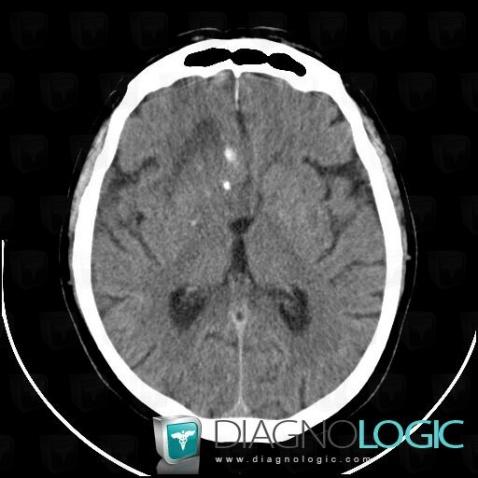
Here is the specific information in the key image above:
- Diagnosis Low grade astrocytoma, Location(s) Ventricles / Periventricular region, with gamuts Intraventricular mass
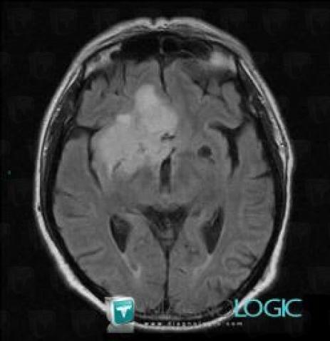
Here is the specific information in the key image above:
- Diagnosis Low grade astrocytoma, Location(s) Cerebral hemispheres, with gamuts Intracerebral T2W or FLAIR hyperintense lesionBasal ganglia and capsule, with gamuts Basal ganglia T2W or FLAIR hyperintense lesionCortico subcortical region, with gamuts Subcortical lesion, Cortical lesionVentricles / Periventricular region, with gamuts Periventricular anomaly seen in MRI
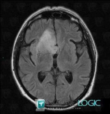
Here is the specific information in the key image above:
- Diagnosis Low grade astrocytoma, Location(s) Ventricles / Periventricular region, with gamuts Intraventricular mass, Ventricular wall nodulesCorpus callosum, with gamuts Corpus callosum lesion
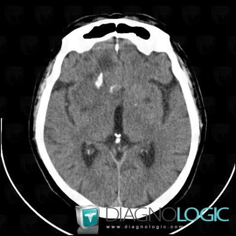
Here is the specific information in the key image above:
- Diagnosis Low grade astrocytoma, Location(s) Basal ganglia and capsule, with gamuts Basal ganglia calcifications, Basal ganglia hypodense lesionCerebral hemispheres, with gamuts Hypodense intracerebral lesion on noncontrast CT, Intracerebral massVentricles / Periventricular region, with gamuts Periventricular hypodensity
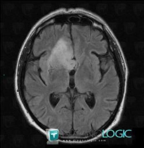
Here is the specific information in the key image above:
- Diagnosis Low grade astrocytoma, Location(s) Ventricles / Periventricular region, with gamuts Intraventricular mass, Ventricular wall nodulesCorpus callosum, with gamuts Corpus callosum lesion
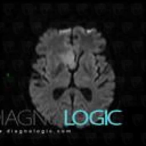
Here is the specific information in the key image above:
- Diagnosis Low grade astrocytoma, Location(s) Cerebral hemispheres, with gamuts DWI hyperintense lesion
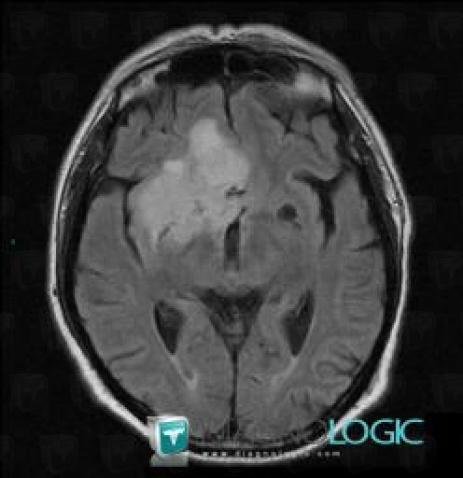
Here is the specific information in the key image above:
- Diagnosis Low grade astrocytoma, Location(s) Cerebral hemispheres, with gamuts Intracerebral T2W or FLAIR hyperintense lesionBasal ganglia and capsule, with gamuts Basal ganglia T2W or FLAIR hyperintense lesionCortico subcortical region, with gamuts Subcortical lesion, Cortical lesionVentricles / Periventricular region, with gamuts Periventricular anomaly seen in MRI
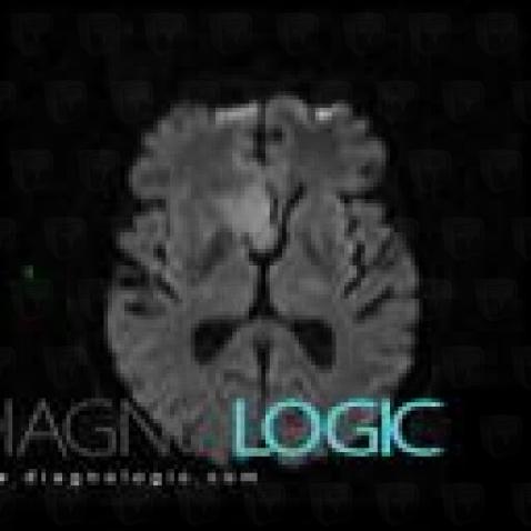
Here is the specific information in the key image above:
- Diagnosis Low grade astrocytoma, Location(s) Cerebral hemispheres, with gamuts DWI hyperintense lesion
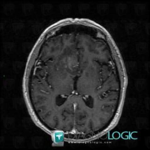
Here is the specific information in the key image above:
- Diagnosis Low grade astrocytoma, Location(s) Cerebral hemispheres, with gamuts Intracerebral lesion with moderate enhancement, Intracerebral mass
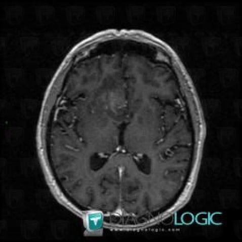
Here is the specific information in the key image above:
- Diagnosis Low grade astrocytoma, Location(s) Cerebral hemispheres, with gamuts Intracerebral lesion with moderate enhancement, Intracerebral mass