The images below illustrate this case for diagnoses Transependymal CSF flow (link to Hydrocephalus), Meningioma, Hydrocephalus, for the modalities (MRI)
Gamuts for this case : :
- Infratentorial T2W or FLAIR hyperintense lesion
- Periventricular anomaly seen in MRI
- Infratentorial extra axial lesion
- Cerebellopontine angle mass
- Focal meningeal enhancement
- Infratentorial lesion with intense enhancement
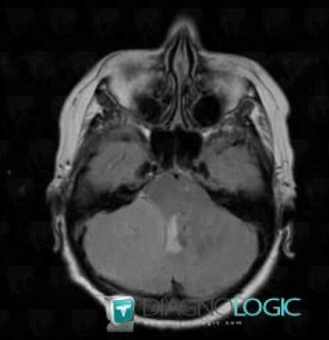
Here is the specific information in the key image above:
- Diagnosis Meningioma, Location(s) Posterior fossa, with gamuts Infratentorial T2W or FLAIR hyperintense lesion
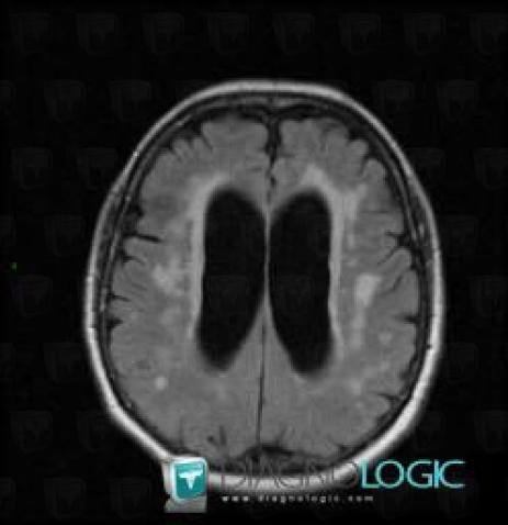
Here is the specific information in the key image above:
- Diagnosis Transependymal CSF flow (link to Hydrocephalus), Location(s) Ventricles / Periventricular region, with gamuts Periventricular anomaly seen in MRI
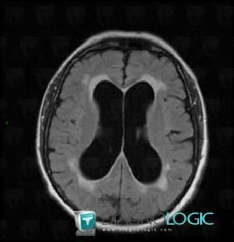
Here is the specific information in the key image above:
- Diagnosis Hydrocephalus, Location(s) Ventricles / Periventricular region, with gamuts Periventricular anomaly seen in MRI
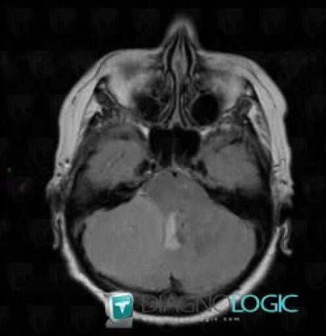
Here is the specific information in the key image above:
- Diagnosis Meningioma, Location(s) Posterior fossa, with gamuts Infratentorial T2W or FLAIR hyperintense lesion
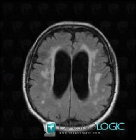
Here is the specific information in the key image above:
- Diagnosis Transependymal CSF flow (link to Hydrocephalus), Location(s) Ventricles / Periventricular region, with gamuts Periventricular anomaly seen in MRI
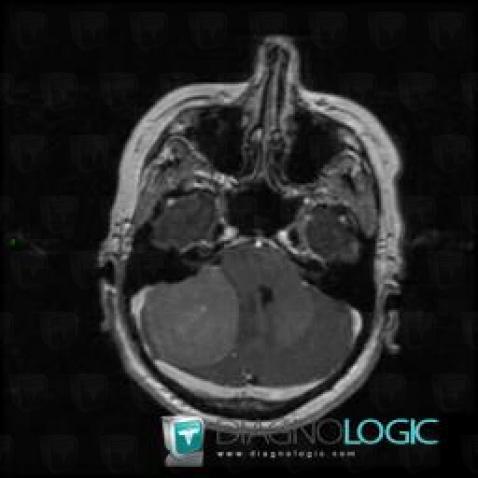
Here is the specific information in the key image above:
- Diagnosis Meningioma, Location(s) Infratentorial peri cerebral spaces, with gamuts Infratentorial extra axial lesion, Focal meningeal enhancementCerebellopontine angle, with gamuts Cerebellopontine angle massPosterior fossa, with gamuts Infratentorial lesion with intense enhancement
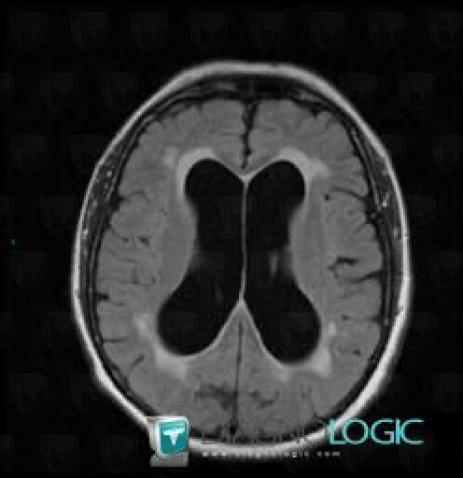
Here is the specific information in the key image above:
- Diagnosis Hydrocephalus, Location(s) Ventricles / Periventricular region, with gamuts Periventricular anomaly seen in MRI
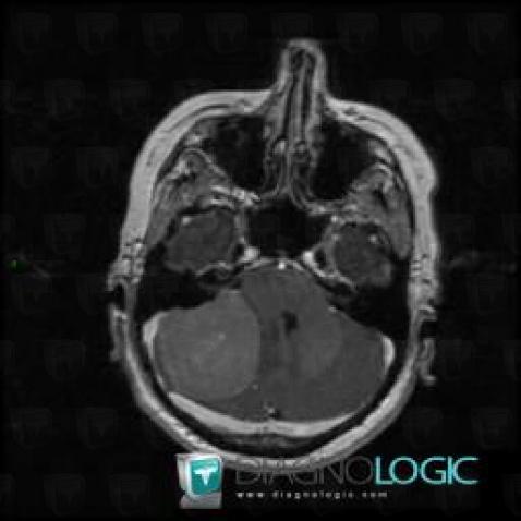
Here is the specific information in the key image above:
- Diagnosis Meningioma, Location(s) Infratentorial peri cerebral spaces, with gamuts Infratentorial extra axial lesion, Focal meningeal enhancementCerebellopontine angle, with gamuts Cerebellopontine angle massPosterior fossa, with gamuts Infratentorial lesion with intense enhancement