The images below illustrate this case for diagnoses Subcapsular hematoma (link to Hemorrhage), Subcapsular hematoma (link to Hematoma), Angiomyolipoma, for the modalities (CT)
Gamuts for this case : :
- Hyperdense renal mass in non enhanced CT
- Lesion in the perinephric space
- Exophytic renal mass
- Solid renal mass
- Fat containing renal mass
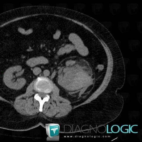
Here is the specific information in the key image above:
- Diagnosis Subcapsular hematoma (link to Hematoma), Location(s) Kidney, with gamuts Hyperdense renal mass in non enhanced CT
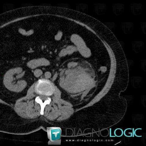
Here is the specific information in the key image above:
- Diagnosis Subcapsular hematoma (link to Hematoma), Location(s) Kidney, with gamuts Hyperdense renal mass in non enhanced CT
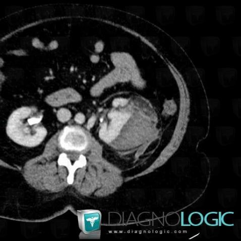
Here is the specific information in the key image above:
- Diagnosis Subcapsular hematoma (link to Hemorrhage), Location(s) Kidney, with gamuts Lesion in the perinephric space
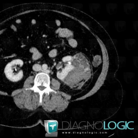
Here is the specific information in the key image above:
- Diagnosis Angiomyolipoma, Location(s) Kidney, with gamuts Exophytic renal mass, Solid renal mass, Fat containing renal mass
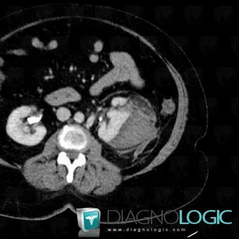
Here is the specific information in the key image above:
- Diagnosis Subcapsular hematoma (link to Hemorrhage), Location(s) Kidney, with gamuts Lesion in the perinephric space
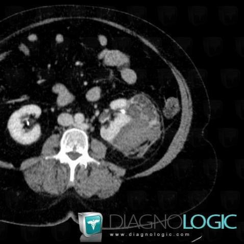
Here is the specific information in the key image above:
- Diagnosis Angiomyolipoma, Location(s) Kidney, with gamuts Exophytic renal mass, Solid renal mass, Fat containing renal mass