The images below illustrate this case for diagnoses Tuberous sclerosis (link to Hamartoma), Hamartoma, Tuberous sclerosis, for the modalities (MRI)
Gamuts for this case : :
- Periventricular anomaly seen in MRI
- Ventricular wall nodules
- Without gamut
- Multifocal intracranial lesions
- Subcortical lesion
- Intracerebral T2W or FLAIR hyperintense lesion
- Cortical lesion
- Parietal posterior or occipital T2WI or FLAIR hyperintense lesion
- Basal ganglia T2W or FLAIR hypointense lesion
- Corpus callosum lesion
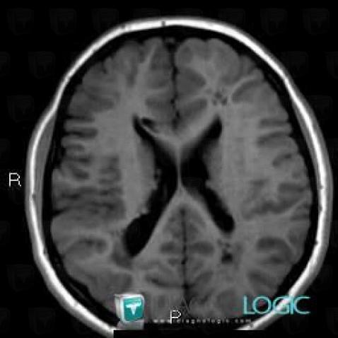
Here is the specific information in the key image above:
- Diagnosis Tuberous sclerosis (link to Hamartoma), Location(s) Ventricles / Periventricular region, with gamuts Periventricular anomaly seen in MRI, Ventricular wall nodules
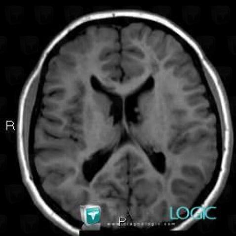
Here is the specific information in the key image above:
- Diagnosis Tuberous sclerosis, Location(s) Ventricles / Periventricular region, with gamuts
- Diagnosis Hamartoma, Location(s) Ventricles / Periventricular region, with gamuts Periventricular anomaly seen in MRI, Ventricular wall nodules
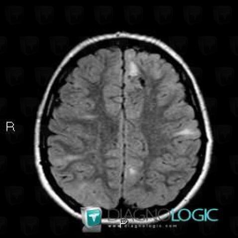
Here is the specific information in the key image above:
- Diagnosis Tuberous sclerosis (link to Hamartoma), Location(s) Cerebral hemispheres, with gamuts Multifocal intracranial lesions, Intracerebral T2W or FLAIR hyperintense lesion
- Diagnosis Hamartoma, Location(s) Cortico subcortical region, with gamuts Subcortical lesion, Cortical lesion
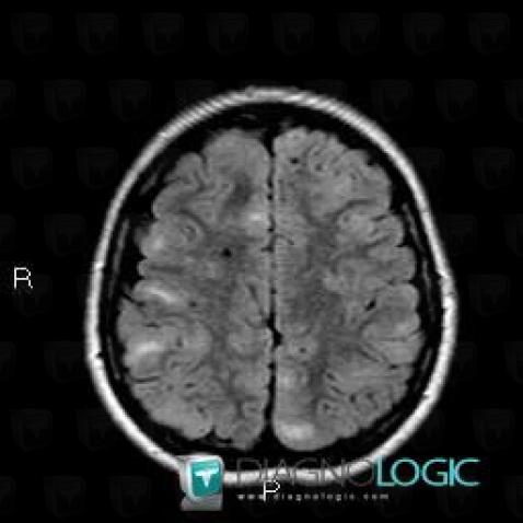
Here is the specific information in the key image above:
- Diagnosis Tuberous sclerosis, Location(s) Cerebral hemispheres, with gamuts Parietal posterior or occipital T2WI or FLAIR hyperintense lesion
- Diagnosis Tuberous sclerosis (link to Hamartoma), Location(s) Cortico subcortical region, with gamuts Cortical lesion, Subcortical lesion
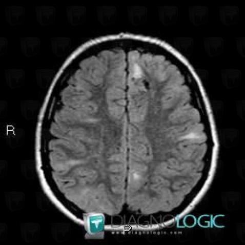
Here is the specific information in the key image above:
- Diagnosis Tuberous sclerosis (link to Hamartoma), Location(s) Cerebral hemispheres, with gamuts Multifocal intracranial lesions, Intracerebral T2W or FLAIR hyperintense lesion
- Diagnosis Hamartoma, Location(s) Cortico subcortical region, with gamuts Subcortical lesion, Cortical lesion
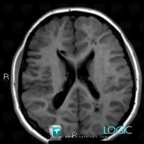
Here is the specific information in the key image above:
- Diagnosis Tuberous sclerosis (link to Hamartoma), Location(s) Ventricles / Periventricular region, with gamuts Periventricular anomaly seen in MRI, Ventricular wall nodules
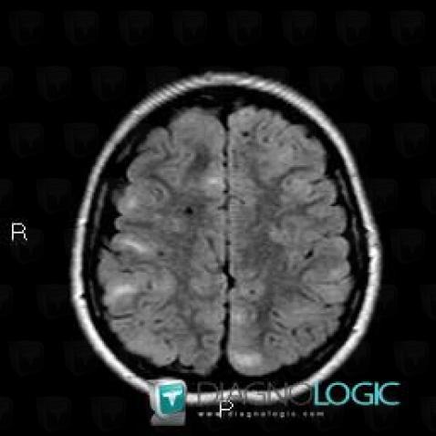
Here is the specific information in the key image above:
- Diagnosis Tuberous sclerosis, Location(s) Cerebral hemispheres, with gamuts Parietal posterior or occipital T2WI or FLAIR hyperintense lesion
- Diagnosis Tuberous sclerosis (link to Hamartoma), Location(s) Cortico subcortical region, with gamuts Cortical lesion, Subcortical lesion
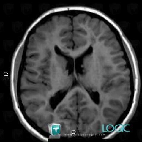
Here is the specific information in the key image above:
- Diagnosis Tuberous sclerosis, Location(s) Ventricles / Periventricular region, with gamuts
- Diagnosis Hamartoma, Location(s) Ventricles / Periventricular region, with gamuts Periventricular anomaly seen in MRI, Ventricular wall nodules
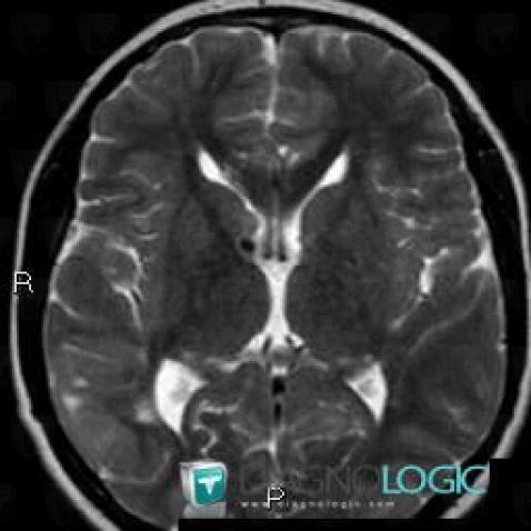
Here is the specific information in the key image above:
- Diagnosis Hamartoma, Location(s) Basal ganglia and capsule, with gamuts Basal ganglia T2W or FLAIR hypointense lesion
- Diagnosis Tuberous sclerosis, Location(s) Basal ganglia and capsule, with gamuts
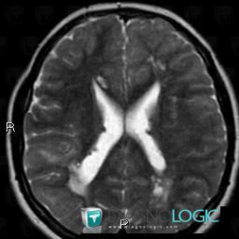
Here is the specific information in the key image above:
- Diagnosis Tuberous sclerosis, Location(s) Corpus callosum, with gamuts Corpus callosum lesion
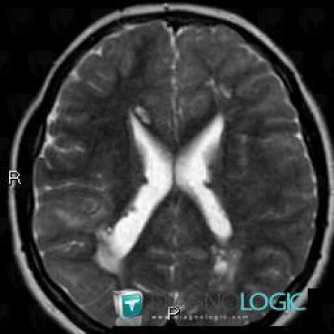
Here is the specific information in the key image above:
- Diagnosis Tuberous sclerosis, Location(s) Corpus callosum, with gamuts Corpus callosum lesion
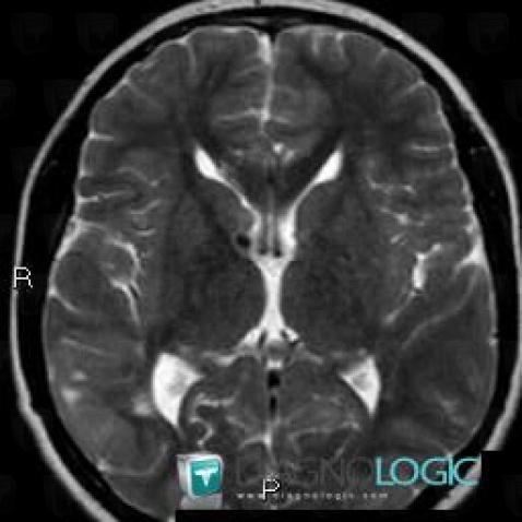
Here is the specific information in the key image above:
- Diagnosis Hamartoma, Location(s) Basal ganglia and capsule, with gamuts Basal ganglia T2W or FLAIR hypointense lesion
- Diagnosis Tuberous sclerosis, Location(s) Basal ganglia and capsule, with gamuts