The images below illustrate this case for diagnoses Radiation therapy lesion (link to Gliosis), Radiation therapy lesion, Phthisis bulbi, Hematoma, for the modalities (CT ,MRI)
Gamuts for this case : :
- Hyperdense intracerebral lesion on noncontrast CT
- Calcified ocular lesion
- Hypodense intracerebral lesion on noncontrast CT
- Basal ganglia hypodense lesion
- Multifocal intracranial lesions
- Temporal lobe T2WI or FLAIR hyperintense lesion
- Intracerebral T2W or FLAIR hyperintense lesion
- White matter disease
- Basal ganglia T2W or FLAIR hyperintense lesion
- Periventricular anomaly seen in MRI
- Without gamut
- Intracerebral T1W hyperintense lesion
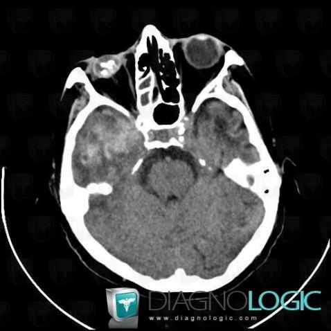
Here is the specific information in the key image above:
- Diagnosis Hematoma, Location(s) Cerebral hemispheres, with gamuts Hyperdense intracerebral lesion on noncontrast CT
- Diagnosis Phthisis bulbi, Location(s) Eye, with gamuts Calcified ocular lesion
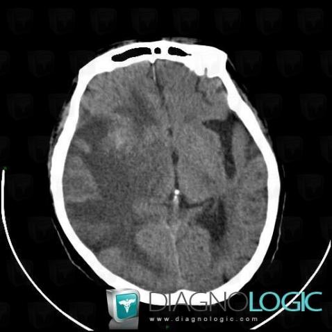
Here is the specific information in the key image above:
- Diagnosis Radiation therapy lesion, Location(s) Cerebral hemispheres, with gamuts Hypodense intracerebral lesion on noncontrast CT, Multifocal intracranial lesionsBasal ganglia and capsule, with gamuts Basal ganglia hypodense lesion
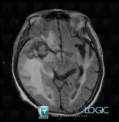
Here is the specific information in the key image above:
- Diagnosis Radiation therapy lesion, Location(s) Cerebral hemispheres, with gamuts Temporal lobe T2WI or FLAIR hyperintense lesion, White matter disease, Multifocal intracranial lesions
- Diagnosis Radiation therapy lesion (link to Gliosis), Location(s) Cerebral hemispheres, with gamuts Intracerebral T2W or FLAIR hyperintense lesion
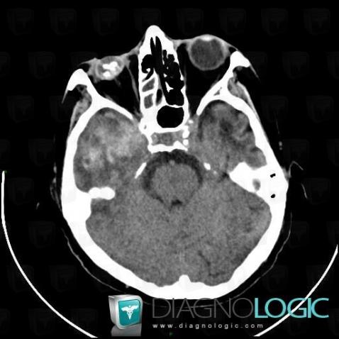
Here is the specific information in the key image above:
- Diagnosis Hematoma, Location(s) Cerebral hemispheres, with gamuts Hyperdense intracerebral lesion on noncontrast CT
- Diagnosis Phthisis bulbi, Location(s) Eye, with gamuts Calcified ocular lesion
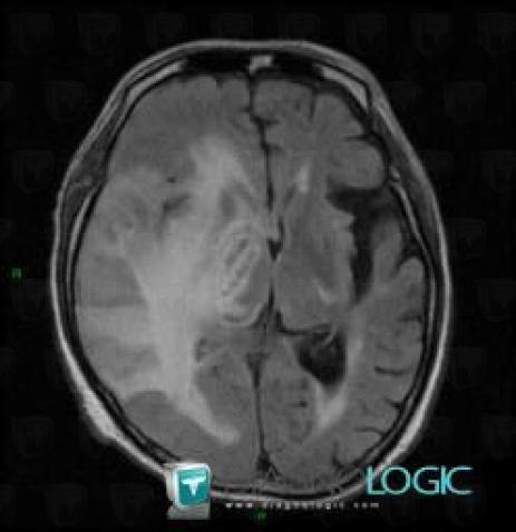
Here is the specific information in the key image above:
- Diagnosis Radiation therapy lesion, Location(s) Basal ganglia and capsule, with gamuts Basal ganglia T2W or FLAIR hyperintense lesion
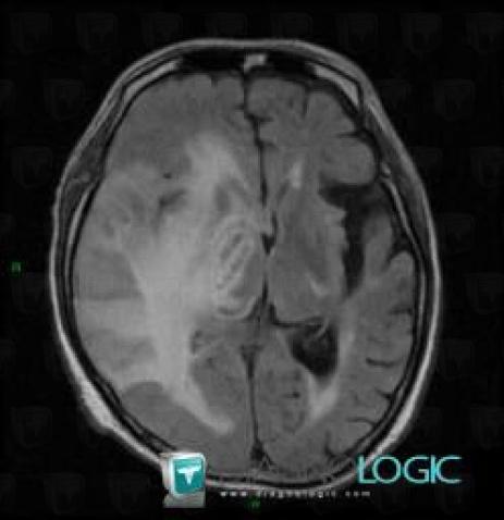
Here is the specific information in the key image above:
- Diagnosis Radiation therapy lesion, Location(s) Basal ganglia and capsule, with gamuts Basal ganglia T2W or FLAIR hyperintense lesion
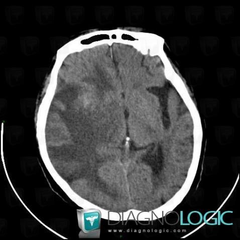
Here is the specific information in the key image above:
- Diagnosis Radiation therapy lesion, Location(s) Cerebral hemispheres, with gamuts Hypodense intracerebral lesion on noncontrast CT, Multifocal intracranial lesionsBasal ganglia and capsule, with gamuts Basal ganglia hypodense lesion
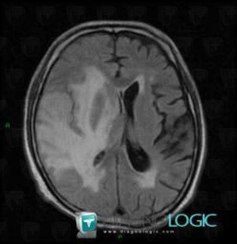
Here is the specific information in the key image above:
- Diagnosis Radiation therapy lesion, Location(s) Ventricles / Periventricular region, with gamuts Periventricular anomaly seen in MRI
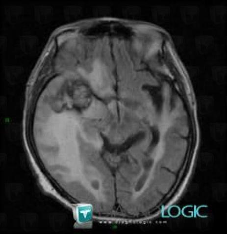
Here is the specific information in the key image above:
- Diagnosis Radiation therapy lesion, Location(s) Cerebral hemispheres, with gamuts Temporal lobe T2WI or FLAIR hyperintense lesion, White matter disease, Multifocal intracranial lesions
- Diagnosis Radiation therapy lesion (link to Gliosis), Location(s) Cerebral hemispheres, with gamuts Intracerebral T2W or FLAIR hyperintense lesion
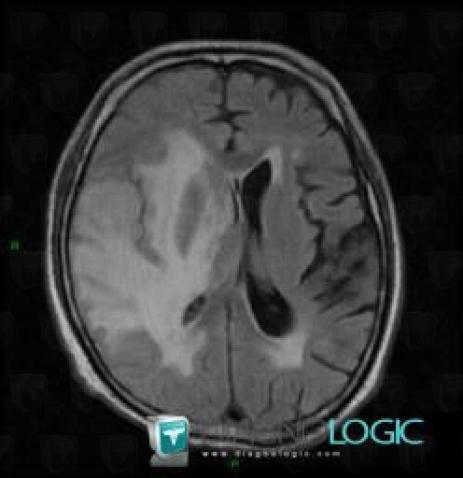
Here is the specific information in the key image above:
- Diagnosis Radiation therapy lesion, Location(s) Ventricles / Periventricular region, with gamuts Periventricular anomaly seen in MRI
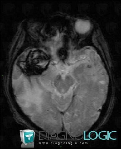
Here is the specific information in the key image above:
- Diagnosis Hematoma, Location(s) Cerebral hemispheres, with gamuts
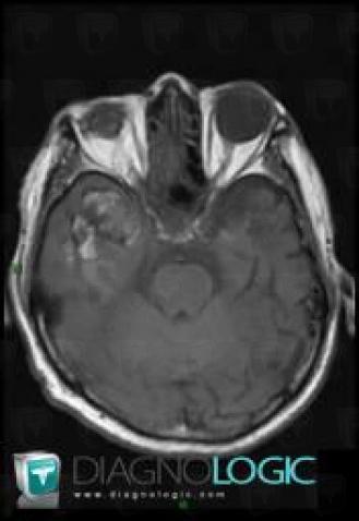
Here is the specific information in the key image above:
- Diagnosis Hematoma, Location(s) Cerebral hemispheres, with gamuts Intracerebral T1W hyperintense lesion
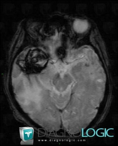
Here is the specific information in the key image above:
- Diagnosis Hematoma, Location(s) Cerebral hemispheres, with gamuts
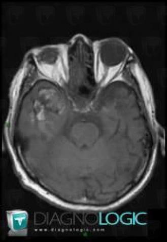
Here is the specific information in the key image above:
- Diagnosis Hematoma, Location(s) Cerebral hemispheres, with gamuts Intracerebral T1W hyperintense lesion



 Patient details - Key images