The images below illustrate this case for diagnoses Fibrous dysplasia, for the modalities (CT ,X rays)
Gamuts for this case : :
- Solitary osteosclerotic bone lesion
- Well-defined osteolysis
- Meta-diaphyseal osteolysis
- Geographic lesion with calcified matrix
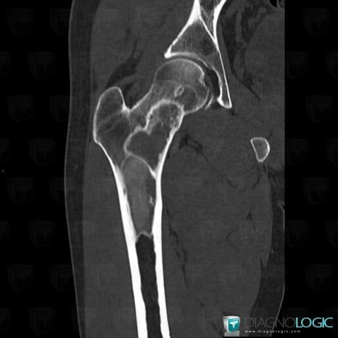
Here is the specific information in the key image above:
- Diagnosis Fibrous dysplasia, Location(s) Femur - Proximal part, with gamuts Well-defined osteolysis, Meta-diaphyseal osteolysis, Solitary osteosclerotic bone lesion
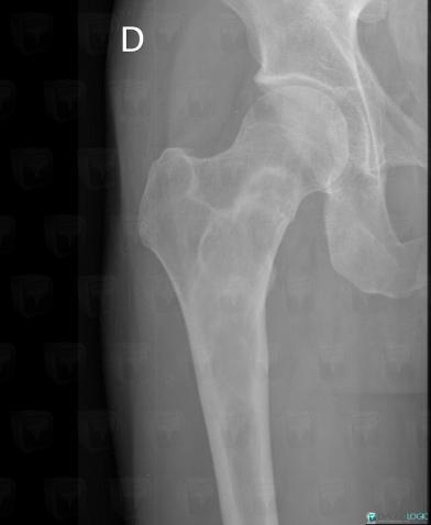
Here is the specific information in the key image above:
- Diagnosis Fibrous dysplasia, Location(s) Femur - Proximal part, with gamuts Solitary osteosclerotic bone lesion, Well-defined osteolysis, Meta-diaphyseal osteolysis, Geographic lesion with calcified matrix
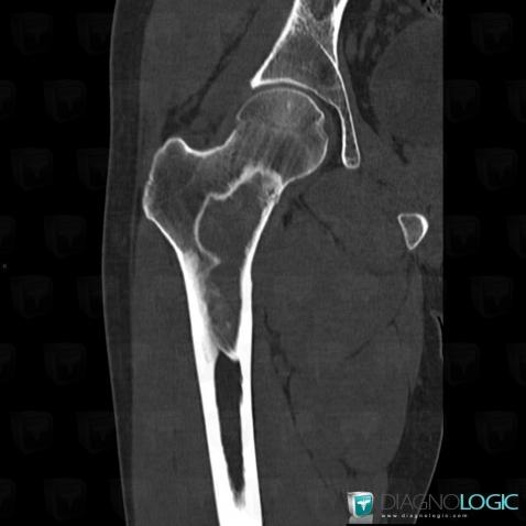
Here is the specific information in the key image above:
- Diagnosis Fibrous dysplasia, Location(s) Femur - Proximal part, with gamuts Geographic lesion with calcified matrix
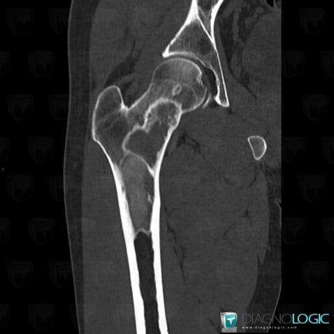
Here is the specific information in the key image above:
- Diagnosis Fibrous dysplasia, Location(s) Femur - Proximal part, with gamuts Well-defined osteolysis, Meta-diaphyseal osteolysis, Solitary osteosclerotic bone lesion
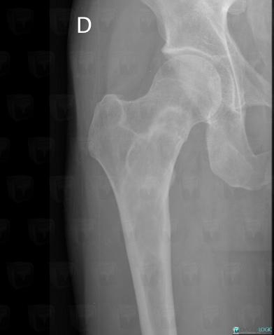
Here is the specific information in the key image above:
- Diagnosis Fibrous dysplasia, Location(s) Femur - Proximal part, with gamuts Solitary osteosclerotic bone lesion, Well-defined osteolysis, Meta-diaphyseal osteolysis