The images below illustrate this case for diagnoses Epidermoid cyst, for the modalities (MRI ,CT)
Gamuts for this case : :
- Hypodense infratentorial lesion on noncontrast CT
- Infratentorial extra axial lesion
- Infratentorial cystic lesion
- DWI hyperintense lesion
- Infratentorial lesion with minimal or no enhancement
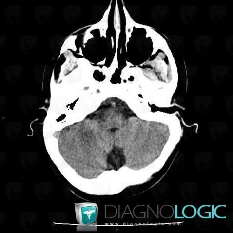
Here is the specific information in the key image above:
- Diagnosis Epidermoid cyst, Location(s) Posterior fossa, with gamuts Hypodense infratentorial lesion on noncontrast CTInfratentorial peri cerebral spaces, with gamuts Infratentorial extra axial lesion
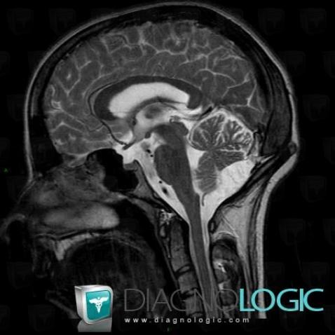
Here is the specific information in the key image above:
- Diagnosis Epidermoid cyst, Location(s) Infratentorial peri cerebral spaces, with gamuts Infratentorial extra axial lesionPosterior fossa, with gamuts Infratentorial cystic lesion
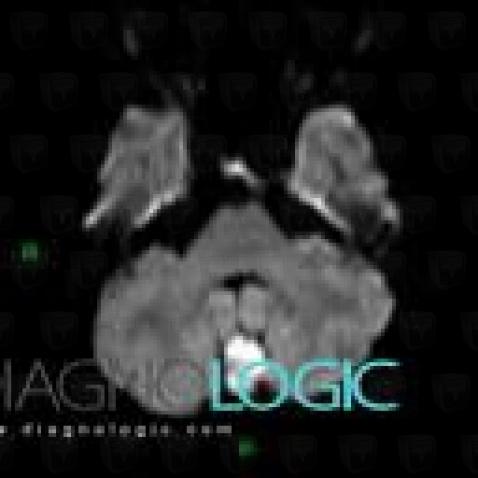
Here is the specific information in the key image above:
- Diagnosis Epidermoid cyst, Location(s) Posterior fossa, with gamuts DWI hyperintense lesion
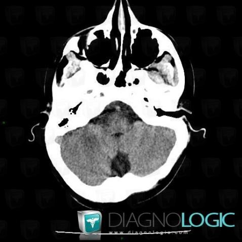
Here is the specific information in the key image above:
- Diagnosis Epidermoid cyst, Location(s) Posterior fossa, with gamuts Hypodense infratentorial lesion on noncontrast CTInfratentorial peri cerebral spaces, with gamuts Infratentorial extra axial lesion
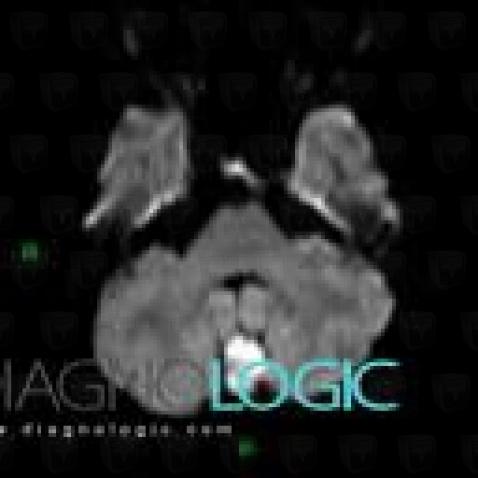
Here is the specific information in the key image above:
- Diagnosis Epidermoid cyst, Location(s) Posterior fossa, with gamuts DWI hyperintense lesion
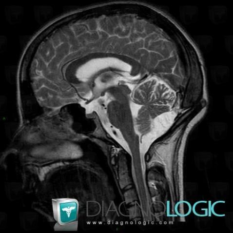
Here is the specific information in the key image above:
- Diagnosis Epidermoid cyst, Location(s) Infratentorial peri cerebral spaces, with gamuts Infratentorial extra axial lesionPosterior fossa, with gamuts Infratentorial cystic lesion
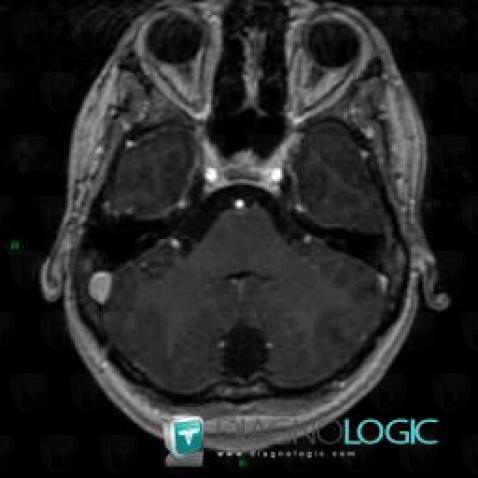
Here is the specific information in the key image above:
- Diagnosis Epidermoid cyst, Location(s) Posterior fossa, with gamuts Infratentorial lesion with minimal or no enhancement
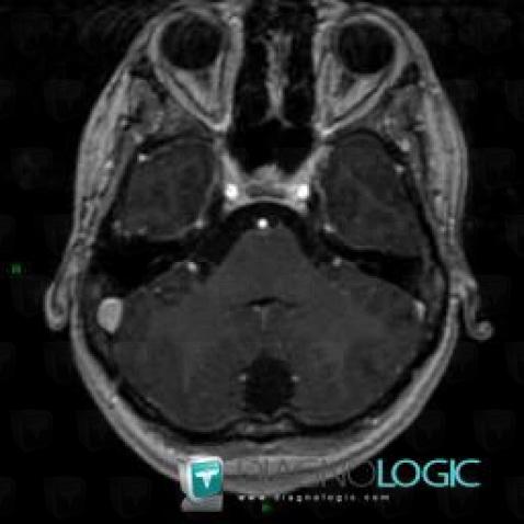
Here is the specific information in the key image above:
- Diagnosis Epidermoid cyst, Location(s) Posterior fossa, with gamuts Infratentorial lesion with minimal or no enhancement