The images below illustrate this case for diagnoses Epidermoid cyst, for the modalities (MRI ,CT)
Gamuts for this case : :
- Infratentorial T2W or FLAIR hypointense lesion
- DWI hyperintense lesion
- Infratentorial T2W or FLAIR hyperintense lesion
- Cerebellopontine angle mass
- Infratentorial lesion with minimal or no enhancement
- Infratentorial T1W hyperintense lesion
- Skull vault thinning
- Osteolytic skull lesion
- Hypodense infratentorial lesion on noncontrast CT
- Intracerebral CSF intensity lesion
- Infratentorial cystic lesion
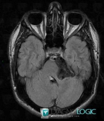
Here is the specific information in the key image above:
- Diagnosis Epidermoid cyst, Location(s) Posterior fossa, with gamuts Infratentorial T2W or FLAIR hypointense lesion
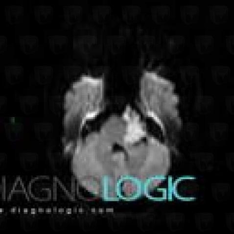
Here is the specific information in the key image above:
- Diagnosis Epidermoid cyst, Location(s) Posterior fossa, with gamuts DWI hyperintense lesion
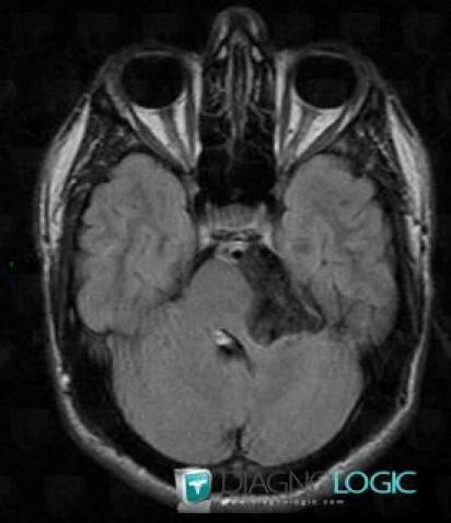
Here is the specific information in the key image above:
- Diagnosis Epidermoid cyst, Location(s) Posterior fossa, with gamuts Infratentorial T2W or FLAIR hypointense lesion
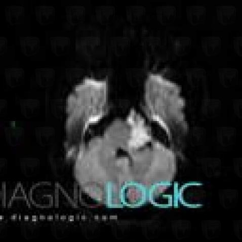
Here is the specific information in the key image above:
- Diagnosis Epidermoid cyst, Location(s) Posterior fossa, with gamuts DWI hyperintense lesion
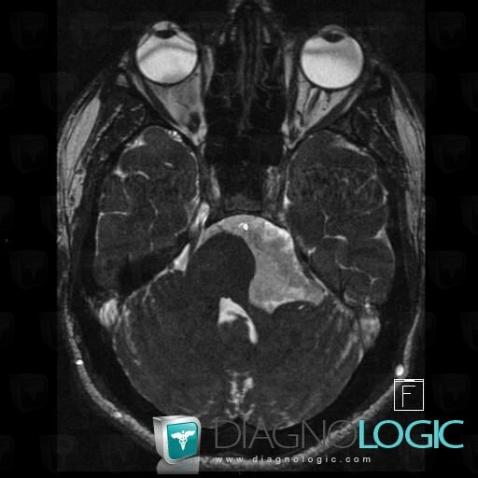
Here is the specific information in the key image above:
- Diagnosis Epidermoid cyst, Location(s) Posterior fossa, with gamuts Infratentorial T2W or FLAIR hyperintense lesionCerebellopontine angle, with gamuts Cerebellopontine angle mass
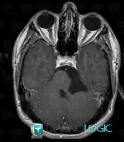
Here is the specific information in the key image above:
- Diagnosis Epidermoid cyst, Location(s) Posterior fossa, with gamuts Infratentorial lesion with minimal or no enhancement
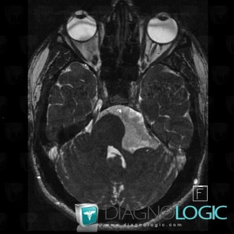
Here is the specific information in the key image above:
- Diagnosis Epidermoid cyst, Location(s) Posterior fossa, with gamuts Infratentorial T2W or FLAIR hyperintense lesionCerebellopontine angle, with gamuts Cerebellopontine angle mass
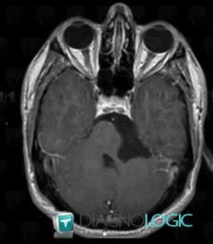
Here is the specific information in the key image above:
- Diagnosis Epidermoid cyst, Location(s) Posterior fossa, with gamuts Infratentorial lesion with minimal or no enhancement
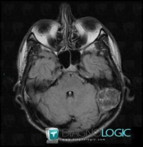
Here is the specific information in the key image above:
- Diagnosis Epidermoid cyst, Location(s) Posterior fossa, with gamuts Infratentorial T2W or FLAIR hypointense lesion

Here is the specific information in the key image above:
- Diagnosis Epidermoid cyst, Location(s) Posterior fossa, with gamuts DWI hyperintense lesion
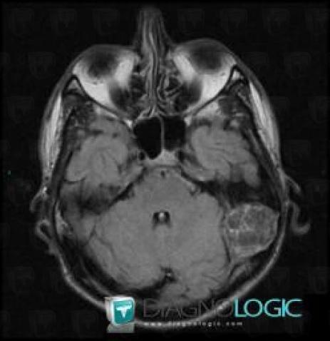
Here is the specific information in the key image above:
- Diagnosis Epidermoid cyst, Location(s) Posterior fossa, with gamuts Infratentorial T2W or FLAIR hypointense lesion

Here is the specific information in the key image above:
- Diagnosis Epidermoid cyst, Location(s) Posterior fossa, with gamuts DWI hyperintense lesion
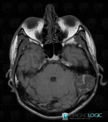
Here is the specific information in the key image above:
- Diagnosis Epidermoid cyst, Location(s) Posterior fossa, with gamuts Infratentorial T1W hyperintense lesion
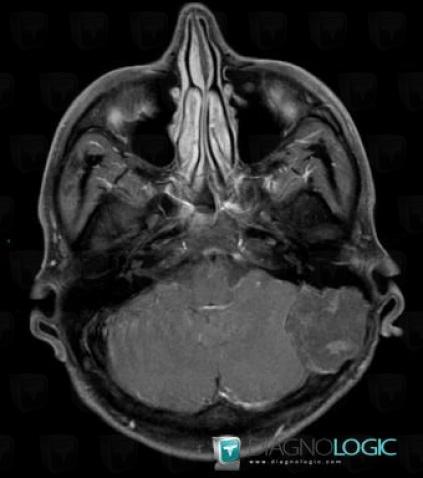
Here is the specific information in the key image above:
- Diagnosis Epidermoid cyst, Location(s) Posterior fossa, with gamuts Infratentorial lesion with minimal or no enhancement
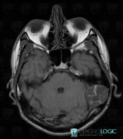
Here is the specific information in the key image above:
- Diagnosis Epidermoid cyst, Location(s) Posterior fossa, with gamuts Infratentorial T1W hyperintense lesion
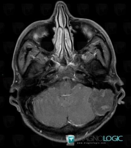
Here is the specific information in the key image above:
- Diagnosis Epidermoid cyst, Location(s) Posterior fossa, with gamuts Infratentorial lesion with minimal or no enhancement
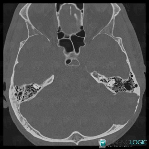
Here is the specific information in the key image above:
- Diagnosis Epidermoid cyst, Location(s) Skull vault, with gamuts Skull vault thinning, Osteolytic skull lesion
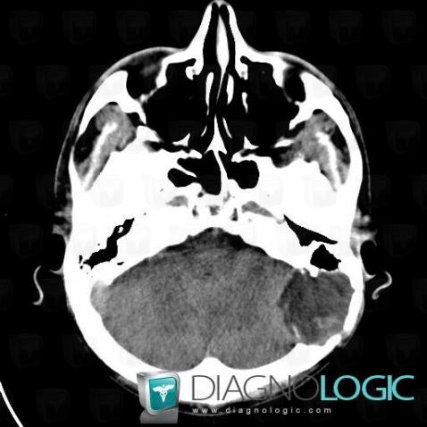
Here is the specific information in the key image above:
- Diagnosis Epidermoid cyst, Location(s) Posterior fossa, with gamuts Hypodense infratentorial lesion on noncontrast CTCerebral hemispheres, with gamuts Intracerebral CSF intensity lesion
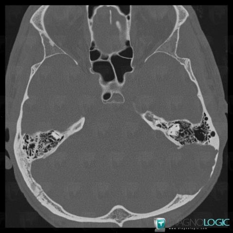
Here is the specific information in the key image above:
- Diagnosis Epidermoid cyst, Location(s) Skull vault, with gamuts Skull vault thinning, Osteolytic skull lesion
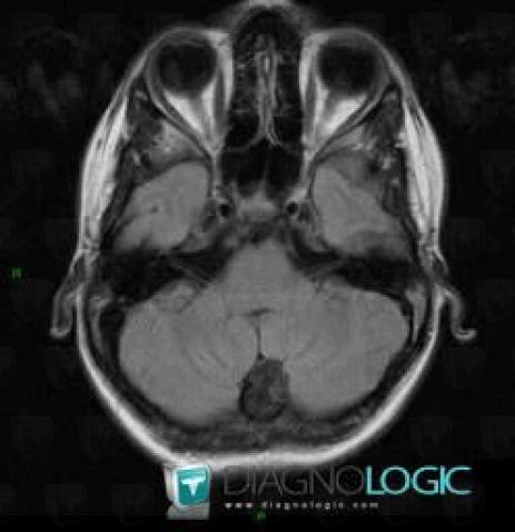
Here is the specific information in the key image above:
- Diagnosis Epidermoid cyst, Location(s) Posterior fossa, with gamuts Infratentorial T2W or FLAIR hypointense lesion
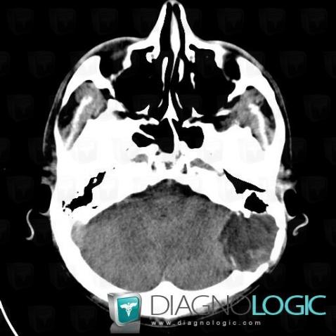
Here is the specific information in the key image above:
- Diagnosis Epidermoid cyst, Location(s) Posterior fossa, with gamuts Hypodense infratentorial lesion on noncontrast CTCerebral hemispheres, with gamuts Intracerebral CSF intensity lesion
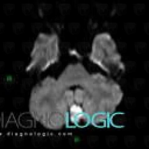
Here is the specific information in the key image above:
- Diagnosis Epidermoid cyst, Location(s) Posterior fossa, with gamuts DWI hyperintense lesion
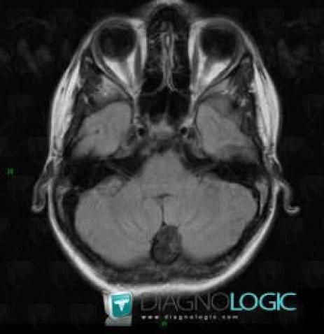
Here is the specific information in the key image above:
- Diagnosis Epidermoid cyst, Location(s) Posterior fossa, with gamuts Infratentorial T2W or FLAIR hypointense lesion
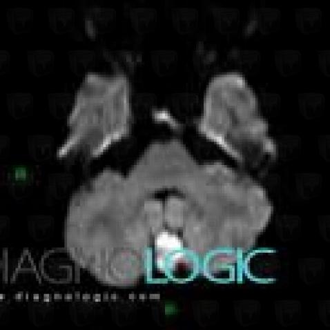
Here is the specific information in the key image above:
- Diagnosis Epidermoid cyst, Location(s) Posterior fossa, with gamuts DWI hyperintense lesion
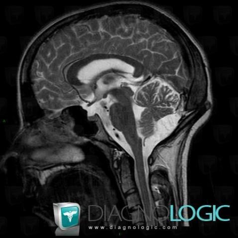
Here is the specific information in the key image above:
- Diagnosis Epidermoid cyst, Location(s) Posterior fossa, with gamuts Infratentorial cystic lesion
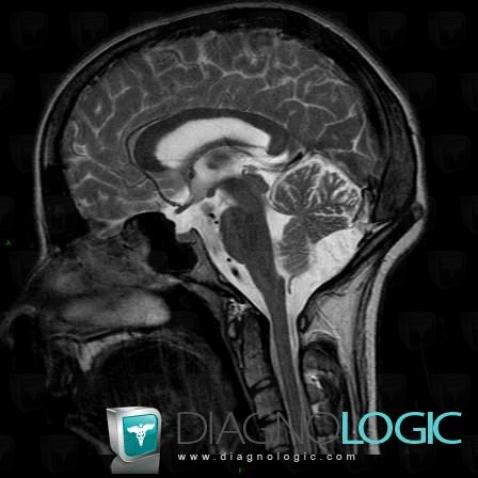
Here is the specific information in the key image above:
- Diagnosis Epidermoid cyst, Location(s) Posterior fossa, with gamuts Infratentorial cystic lesion
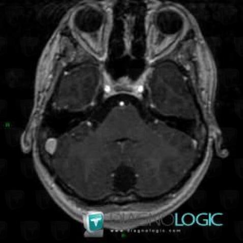
Here is the specific information in the key image above:
- Diagnosis Epidermoid cyst, Location(s) Posterior fossa, with gamuts Infratentorial lesion with minimal or no enhancement
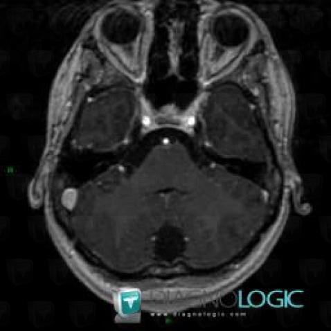
Here is the specific information in the key image above:
- Diagnosis Epidermoid cyst, Location(s) Posterior fossa, with gamuts Infratentorial lesion with minimal or no enhancement