The images below illustrate this case for diagnoses Ependymoma, for the modalities (MRI)
Gamuts for this case : :
- Intraventricular mass
- Infratentorial T2W or FLAIR hyperintense lesion
- Infratentorial cystic lesion
- Infratentorial lesion with intense enhancement
- Infratentorial lesion with ring enhancement
- Ventricular wall nodules
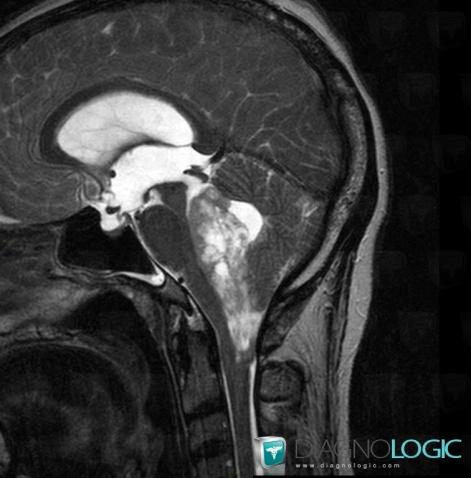
Here is the specific information in the key image above:
- Diagnosis Ependymoma, Location(s) V4 and vermis, with gamuts Intraventricular massPosterior fossa, with gamuts Infratentorial T2W or FLAIR hyperintense lesion
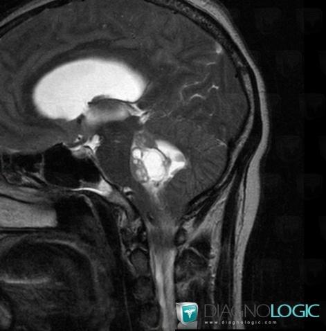
Here is the specific information in the key image above:
- Diagnosis Ependymoma, Location(s) Posterior fossa, with gamuts Infratentorial cystic lesion
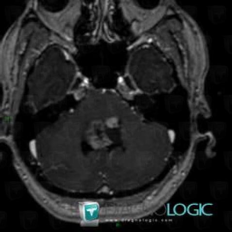
Here is the specific information in the key image above:
- Diagnosis Ependymoma, Location(s) Posterior fossa, with gamuts Infratentorial lesion with intense enhancement, Infratentorial lesion with ring enhancement
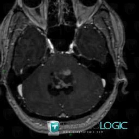
Here is the specific information in the key image above:
- Diagnosis Ependymoma, Location(s) V4 and vermis, with gamuts Ventricular wall nodules
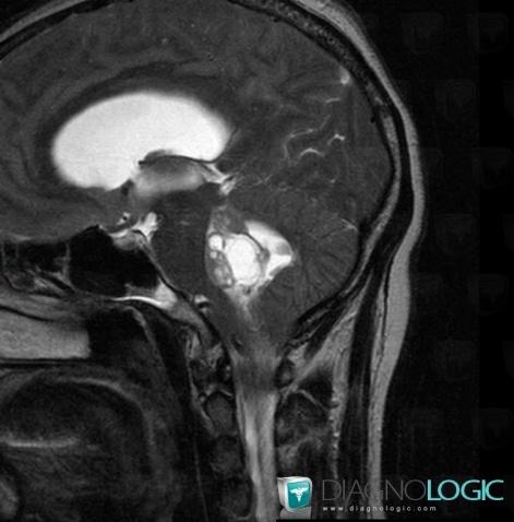
Here is the specific information in the key image above:
- Diagnosis Ependymoma, Location(s) Posterior fossa, with gamuts Infratentorial cystic lesion
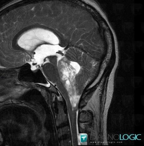
Here is the specific information in the key image above:
- Diagnosis Ependymoma, Location(s) V4 and vermis, with gamuts Intraventricular massPosterior fossa, with gamuts Infratentorial T2W or FLAIR hyperintense lesion
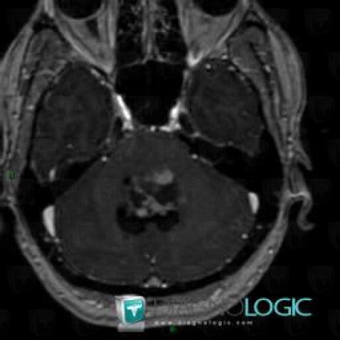
Here is the specific information in the key image above:
- Diagnosis Ependymoma, Location(s) V4 and vermis, with gamuts Ventricular wall nodules
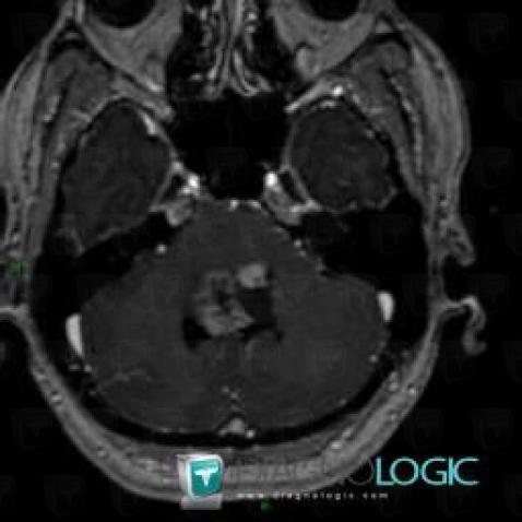
Here is the specific information in the key image above:
- Diagnosis Ependymoma, Location(s) Posterior fossa, with gamuts Infratentorial lesion with intense enhancement, Infratentorial lesion with ring enhancement