The images below illustrate this case for diagnoses Encephalocele, for the modalities (CT ,MRI)
Gamuts for this case : :
- Middle ear or mastoid osteolysis
- Middle ear lesion
- Congenital malformation of the brain
- Without gamut
- Temporal bone osteolysis or demineralisation
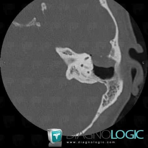
Here is the specific information in the key image above:
- Diagnosis Encephalocele, Location(s) Middle ear, with gamuts Middle ear or mastoid osteolysis, Middle ear lesion
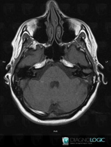
Here is the specific information in the key image above:
- Diagnosis Encephalocele, Location(s) Cerebral hemispheres, with gamuts Congenital malformation of the brain
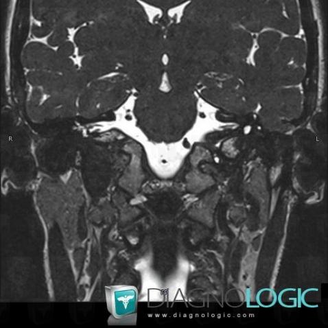
Here is the specific information in the key image above:
- Diagnosis Encephalocele, Location(s) Temporal bone, with gamuts Middle ear, with gamuts Middle ear lesion
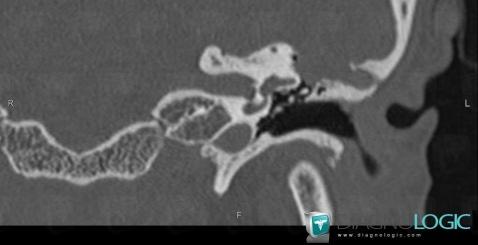
Here is the specific information in the key image above:
- Diagnosis Encephalocele, Location(s) Temporal bone, with gamuts Middle ear or mastoid osteolysis, Temporal bone osteolysis or demineralisation
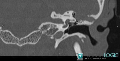
Here is the specific information in the key image above:
- Diagnosis Encephalocele, Location(s) Cerebral hemispheres, with gamuts Congenital malformation of the brain