The images below illustrate this case for diagnoses Paraseptal emphysema (link to Emphysema), Scleroderma (link to NSIP), Scleroderma (link to Pulmonary fibrosis), Scleroderma, for the modalities (CT)
Gamuts for this case : :
- Multiple cystic lung lesions
- Diffuse esophageal dilatation
- Pulmonary fibrosis
- Large symetric area of ground glass opacity
- Lower lung zone disease
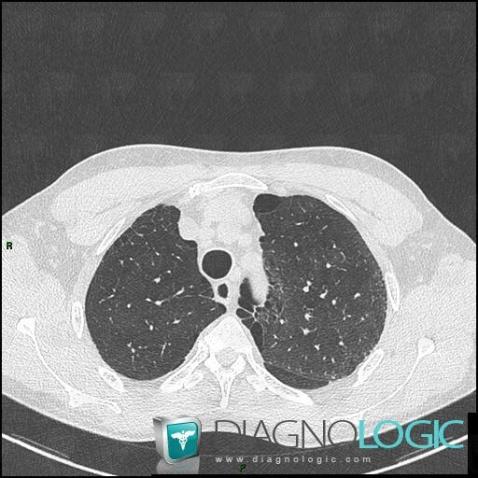
Here is the specific information in the key image above:
- Diagnosis Paraseptal emphysema (link to Emphysema), Location(s) Pulmonary parenchyma, with gamuts Multiple cystic lung lesions
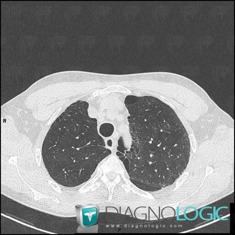
Here is the specific information in the key image above:
- Diagnosis Paraseptal emphysema (link to Emphysema), Location(s) Pulmonary parenchyma, with gamuts Multiple cystic lung lesions
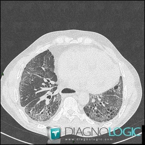
Here is the specific information in the key image above:
- Diagnosis Scleroderma, Location(s) Oesophagus, with gamuts Diffuse esophageal dilatation
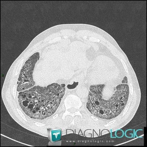
Here is the specific information in the key image above:
- Diagnosis Scleroderma, Location(s) Pulmonary parenchyma, with gamuts Pulmonary fibrosis, Lower lung zone disease
- Diagnosis Scleroderma (link to NSIP), Location(s) Pulmonary parenchyma, with gamuts Large symetric area of ground glass opacity
- Diagnosis Scleroderma (link to Pulmonary fibrosis), Location(s) Pulmonary parenchyma, with gamuts Multiple cystic lung lesions
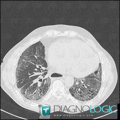
Here is the specific information in the key image above:
- Diagnosis Scleroderma, Location(s) Oesophagus, with gamuts Diffuse esophageal dilatation
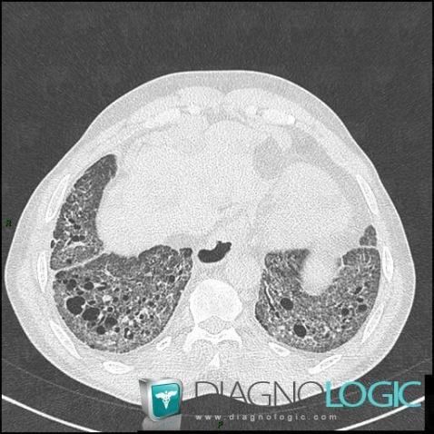
Here is the specific information in the key image above:
- Diagnosis Scleroderma, Location(s) Pulmonary parenchyma, with gamuts Pulmonary fibrosis, Lower lung zone disease
- Diagnosis Scleroderma (link to NSIP), Location(s) Pulmonary parenchyma, with gamuts Large symetric area of ground glass opacity
- Diagnosis Scleroderma (link to Pulmonary fibrosis), Location(s) Pulmonary parenchyma, with gamuts Multiple cystic lung lesions