The images below illustrate this case for diagnoses Centrilobular emphysema (link to Emphysema), Bronchogenic carcinoma (link to Obstructive pneumonia), Metastasis, Bronchogenic carcinoma, Intra alveolar hemorrhage, Venous compression, Lymphatic obstruction, for the modalities (CT)
Gamuts for this case : :
- Multiple pulmonary nodule
- Multiple cystic lung lesions
- Focal area of ground glass opacity
- Smooth peribronchovacular / interlobular thickening
- Peribronchovacular / interlobular thickening
- Localized consolidation
- Solitary pulmonary mass
- Chronic consolidation
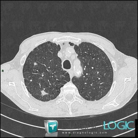
Here is the specific information in the key image above:
- Diagnosis Metastasis, Location(s) Pulmonary parenchyma, with gamuts Multiple pulmonary nodule
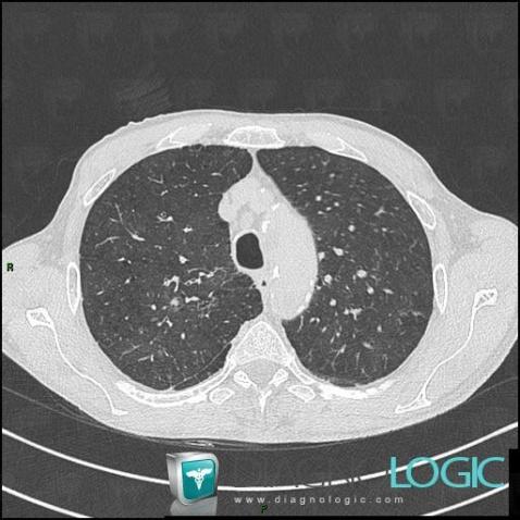
Here is the specific information in the key image above:
- Diagnosis Centrilobular emphysema (link to Emphysema), Location(s) Pulmonary parenchyma, with gamuts Multiple cystic lung lesions
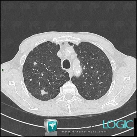
Here is the specific information in the key image above:
- Diagnosis Metastasis, Location(s) Pulmonary parenchyma, with gamuts Multiple pulmonary nodule
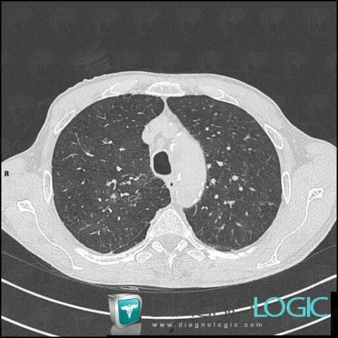
Here is the specific information in the key image above:
- Diagnosis Centrilobular emphysema (link to Emphysema), Location(s) Pulmonary parenchyma, with gamuts Multiple cystic lung lesions
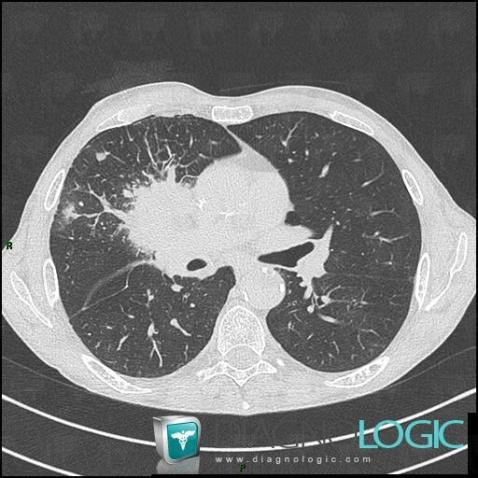
Here is the specific information in the key image above:
- Diagnosis Intra alveolar hemorrhage, Location(s) Pulmonary parenchyma, with gamuts Focal area of ground glass opacity
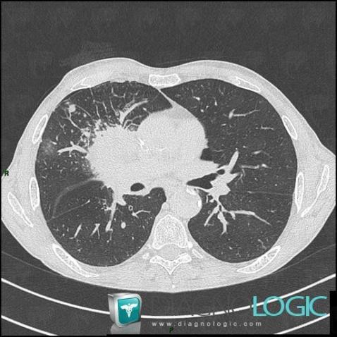
Here is the specific information in the key image above:
- Diagnosis Lymphatic obstruction, Location(s) Pulmonary parenchyma, with gamuts Smooth peribronchovacular / interlobular thickening, Peribronchovacular / interlobular thickening
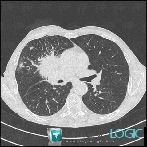
Here is the specific information in the key image above:
- Diagnosis Intra alveolar hemorrhage, Location(s) Pulmonary parenchyma, with gamuts Focal area of ground glass opacity
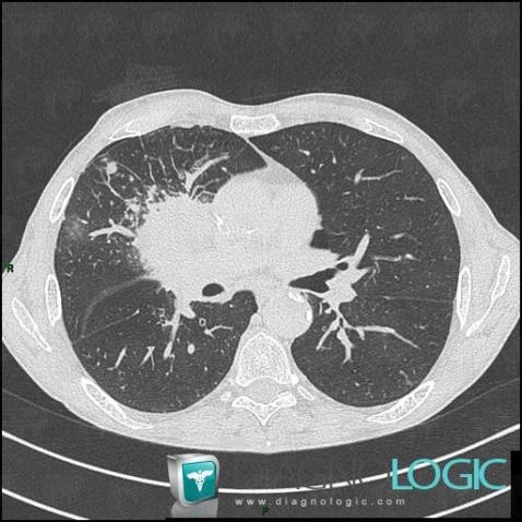
Here is the specific information in the key image above:
- Diagnosis Lymphatic obstruction, Location(s) Pulmonary parenchyma, with gamuts Smooth peribronchovacular / interlobular thickening, Peribronchovacular / interlobular thickening
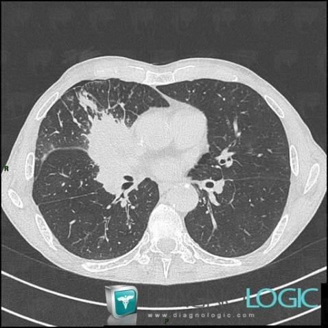
Here is the specific information in the key image above:
- Diagnosis Bronchogenic carcinoma (link to Obstructive pneumonia), Location(s) Pulmonary parenchyma, with gamuts Localized consolidation, Chronic consolidation
- Diagnosis Bronchogenic carcinoma, Location(s) Pulmonary parenchyma, with gamuts Solitary pulmonary mass
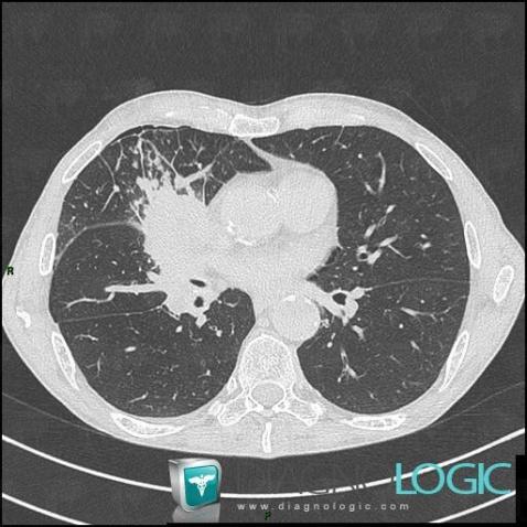
Here is the specific information in the key image above:
- Diagnosis Venous compression, Location(s) Pulmonary parenchyma, with gamuts Smooth peribronchovacular / interlobular thickening, Peribronchovacular / interlobular thickening
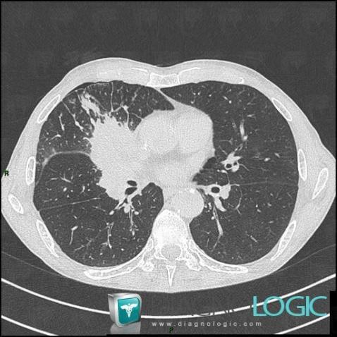
Here is the specific information in the key image above:
- Diagnosis Bronchogenic carcinoma (link to Obstructive pneumonia), Location(s) Pulmonary parenchyma, with gamuts Localized consolidation, Chronic consolidation
- Diagnosis Bronchogenic carcinoma, Location(s) Pulmonary parenchyma, with gamuts Solitary pulmonary mass
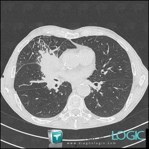
Here is the specific information in the key image above:
- Diagnosis Venous compression, Location(s) Pulmonary parenchyma, with gamuts Smooth peribronchovacular / interlobular thickening, Peribronchovacular / interlobular thickening