The images below illustrate this case for diagnoses Subchondral cyst (link to Degenerative joint disease), CPPD (link to Chondrocalcinosis), Renal osteodystrophy (link to Hyperparathyroidism), Renal osteodystrophy, Subchondral cyst, for the modalities (CT)
Gamuts for this case : :
- Parosteal erosion
- Cortical osteolysis
- Sub periosteal erosion
- Intraarticular or periarticular calcification
- Epiphyseal osteolysis
- Well-defined osteolysis
- Bilateral symmetrical sacroiliac disease
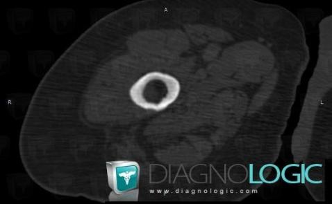
Here is the specific information in the key image above:
- Diagnosis Renal osteodystrophy (link to Hyperparathyroidism), Location(s) Femur - Mid part, with gamuts Parosteal erosion, Cortical osteolysis, Sub periosteal erosion
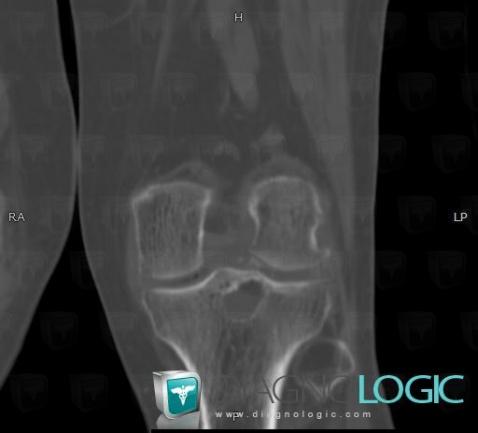
Here is the specific information in the key image above:
- Diagnosis CPPD (link to Chondrocalcinosis), Location(s) Femorotibial joints - Intercondylar notch, with gamuts Intraarticular or periarticular calcification
- Diagnosis Subchondral cyst (link to Degenerative joint disease), Location(s) Tibia - Proximal part, with gamuts Epiphyseal osteolysis
- Diagnosis Subchondral cyst, Location(s) Tibia - Proximal part, with gamuts Well-defined osteolysis
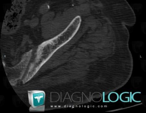
Here is the specific information in the key image above:
- Diagnosis Renal osteodystrophy, Location(s) Sacro iliac joint, with gamuts Bilateral symmetrical sacroiliac disease
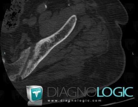
Here is the specific information in the key image above:
- Diagnosis Renal osteodystrophy, Location(s)
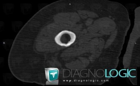
Here is the specific information in the key image above:
- Diagnosis Renal osteodystrophy (link to Hyperparathyroidism), Location(s) Femur - Mid part, with gamuts Parosteal erosion
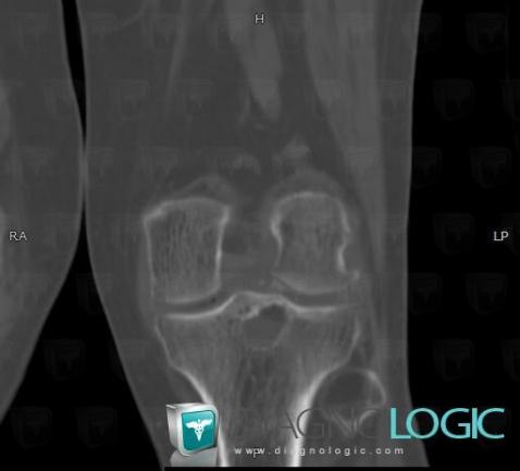
Here is the specific information in the key image above:
- Diagnosis CPPD (link to Chondrocalcinosis), Location(s) Femorotibial joints - Intercondylar notch, with gamuts Intraarticular or periarticular calcification
- Diagnosis Subchondral cyst (link to Degenerative joint disease), Location(s) Tibia - Proximal part, with gamuts
- Diagnosis Subchondral cyst, Location(s) Tibia - Proximal part, with gamuts Well-defined osteolysis
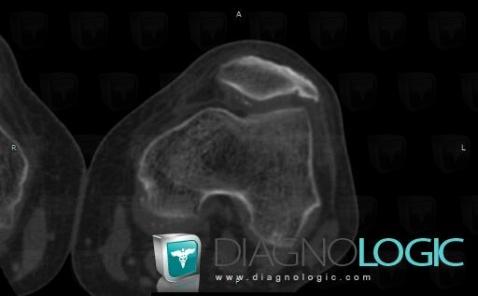
Here is the specific information in the key image above:
- Diagnosis CPPD (link to Chondrocalcinosis), Location(s) Patellofemoral joint, with gamuts