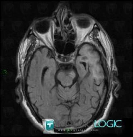
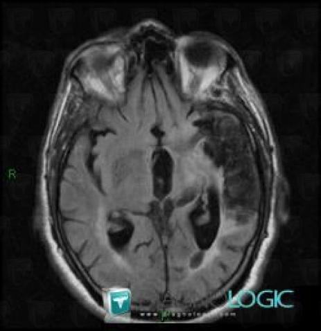
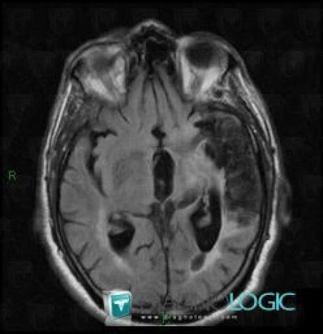
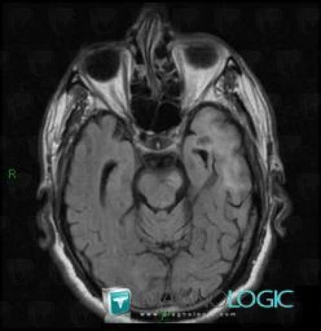
The images below illustrate this case for diagnoses Cerebral infarction, Wallerian degeneration, for the modalities (MRI)
Gamuts for this case : :
- Brainstem lesion
- Brainstem Hyperintense T2WI or FLAIR lesion
- Without gamut

Here is the specific information in the key image above:
- Diagnosis Wallerian degeneration, Location(s) Brainstem, with gamuts Brainstem lesion, Brainstem Hyperintense T2WI or FLAIR lesion

Here is the specific information in the key image above:
- Diagnosis Cerebral infarction, Location(s) Cerebral hemispheres, with gamuts

Here is the specific information in the key image above:
- Diagnosis Cerebral infarction, Location(s) Cerebral hemispheres, with gamuts

Here is the specific information in the key image above:
- Diagnosis Wallerian degeneration, Location(s) Brainstem, with gamuts Brainstem lesion, Brainstem Hyperintense T2WI or FLAIR lesion