The images below illustrate this case for diagnoses Small cell lung carcinoma (link to Bronchogenic carcinoma), Bronchogenic carcinoma (link to Obstructive pneumonia), Bronchogenic carcinoma (link to Tracheal cancer), Metastasis, Bronchogenic carcinoma, for the modalities (US ,CT ,X rays)
Gamuts for this case : :
- Hyperechoic hepatic lesion
- Middle mediastinal mass
- Posterior mediastinal mass
- Low density in portal phase hepatic lesion
- Solid hepatic lesion
- Localized consolidation
- Pulmonary mass with hilar attachment
- Solitary pulmonary mass
- Chronic consolidation
- Hilar mass
- Unilateral hilar enlargement
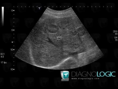
Here is the specific information in the key image above:
- Diagnosis Metastasis, Location(s) Liver, with gamuts Hyperechoic hepatic lesion
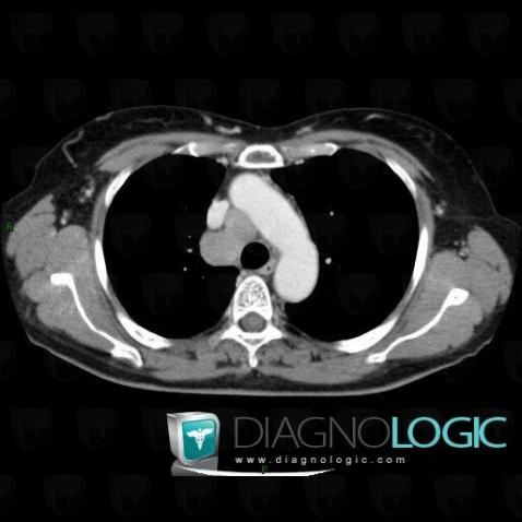
Here is the specific information in the key image above:
- Diagnosis Metastasis, Location(s) Mediastinum, with gamuts Middle mediastinal mass
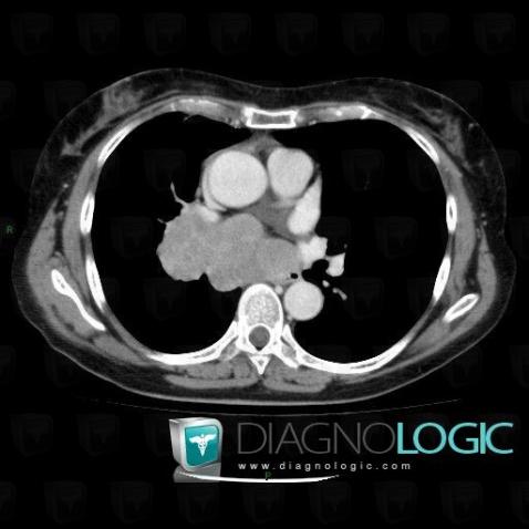
Here is the specific information in the key image above:
- Diagnosis Bronchogenic carcinoma (link to Tracheal cancer), Location(s) Mediastinum, with gamuts Middle mediastinal mass
- Diagnosis Metastasis, Location(s) Mediastinum, with gamuts Posterior mediastinal mass
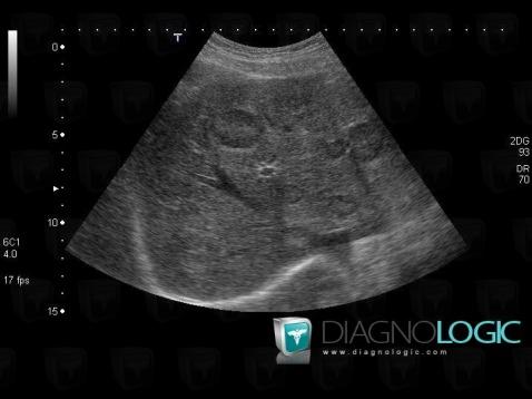
Here is the specific information in the key image above:
- Diagnosis Metastasis, Location(s) Liver, with gamuts Hyperechoic hepatic lesion
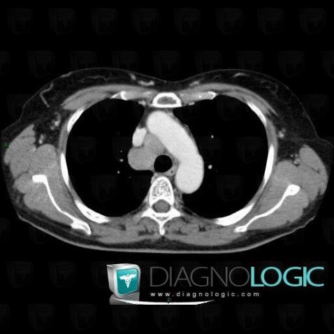
Here is the specific information in the key image above:
- Diagnosis Metastasis, Location(s) Mediastinum, with gamuts Middle mediastinal mass
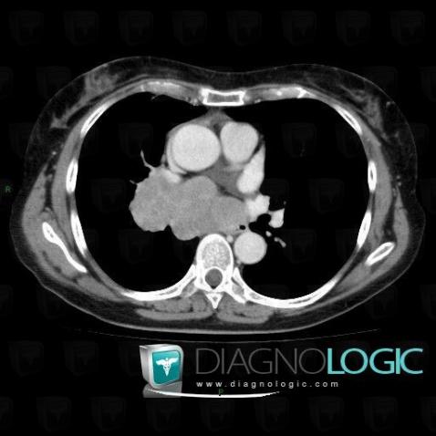
Here is the specific information in the key image above:
- Diagnosis Bronchogenic carcinoma (link to Tracheal cancer), Location(s) Mediastinum, with gamuts Middle mediastinal mass
- Diagnosis Metastasis, Location(s) Mediastinum, with gamuts Posterior mediastinal mass
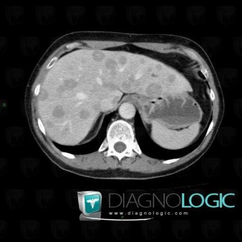
Here is the specific information in the key image above:
- Diagnosis Metastasis, Location(s) Liver, with gamuts Low density in portal phase hepatic lesion, Solid hepatic lesion
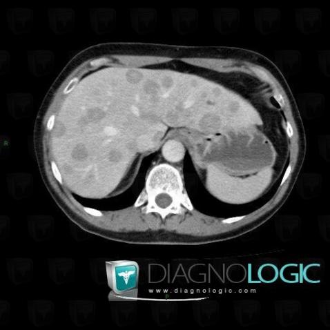
Here is the specific information in the key image above:
- Diagnosis Metastasis, Location(s) Liver, with gamuts Low density in portal phase hepatic lesion, Solid hepatic lesion
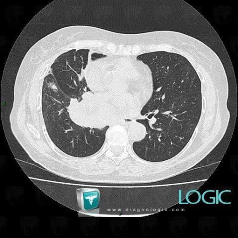
Here is the specific information in the key image above:
- Diagnosis Bronchogenic carcinoma (link to Obstructive pneumonia), Location(s) Pulmonary parenchyma, with gamuts Localized consolidation, Chronic consolidation
- Diagnosis Bronchogenic carcinoma, Location(s) Pulmonary parenchyma, with gamuts Pulmonary mass with hilar attachment, Solitary pulmonary mass
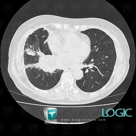
Here is the specific information in the key image above:
- Diagnosis Small cell lung carcinoma (link to Bronchogenic carcinoma), Location(s) Pulmonary parenchyma, with gamuts Solitary pulmonary mass
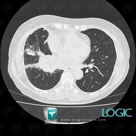
Here is the specific information in the key image above:
- Diagnosis Small cell lung carcinoma (link to Bronchogenic carcinoma), Location(s) Pulmonary parenchyma, with gamuts
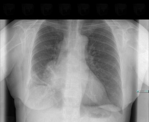
Here is the specific information in the key image above:
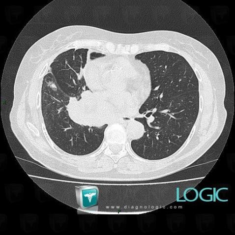
Here is the specific information in the key image above:
- Diagnosis Bronchogenic carcinoma (link to Obstructive pneumonia), Location(s) Pulmonary parenchyma, with gamuts Localized consolidation, Chronic consolidation
- Diagnosis Bronchogenic carcinoma, Location(s) Pulmonary parenchyma, with gamuts Pulmonary mass with hilar attachment, Solitary pulmonary mass
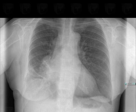
Here is the specific information in the key image above:
- Diagnosis Bronchogenic carcinoma, Location(s) Hila, with gamuts Hilar mass, Unilateral hilar enlargementPulmonary parenchyma, with gamuts Pulmonary mass with hilar attachment, Solitary pulmonary mass
- Diagnosis Bronchogenic carcinoma (link to Obstructive pneumonia), Location(s) Pulmonary parenchyma, with gamuts Chronic consolidation, Localized consolidation