The images below illustrate this case for diagnoses Arteriovenous malformation, for the modalities (MRI)
Gamuts for this case : :
- Intracerebral T2W or FLAIR hypointense lesion
- Subcortical lesion
- Cortical lesion
- Gyriform anomaly
- Intracerebral lesion with intense enhancement
- Multifocal intracranial lesions
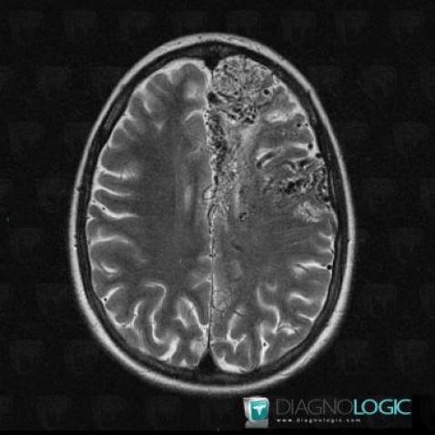
Here is the specific information in the key image above:
- Diagnosis Arteriovenous malformation, Location(s) Cerebral hemispheres, with gamuts Intracerebral T2W or FLAIR hypointense lesion
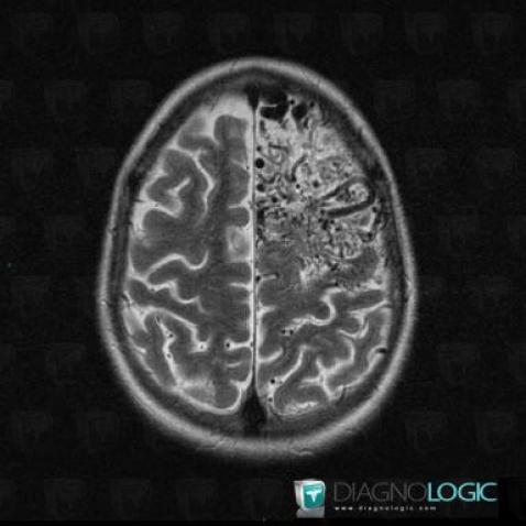
Here is the specific information in the key image above:
- Diagnosis Arteriovenous malformation, Location(s) Cortico subcortical region, with gamuts Subcortical lesion, Cortical lesion
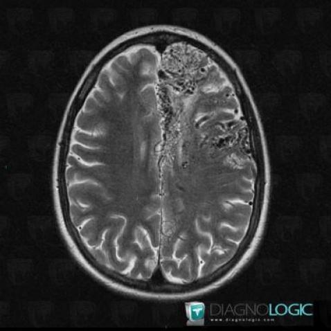
Here is the specific information in the key image above:
- Diagnosis Arteriovenous malformation, Location(s) Cerebral hemispheres, with gamuts Intracerebral T2W or FLAIR hypointense lesion
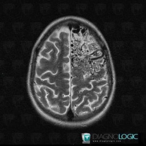
Here is the specific information in the key image above:
- Diagnosis Arteriovenous malformation, Location(s) Cortico subcortical region, with gamuts Subcortical lesion, Cortical lesion
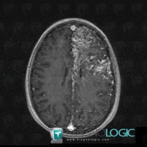
Here is the specific information in the key image above:
- Diagnosis Arteriovenous malformation, Location(s) Cortico subcortical region, with gamuts Gyriform anomalyCerebral hemispheres, with gamuts Intracerebral lesion with intense enhancement, Multifocal intracranial lesions
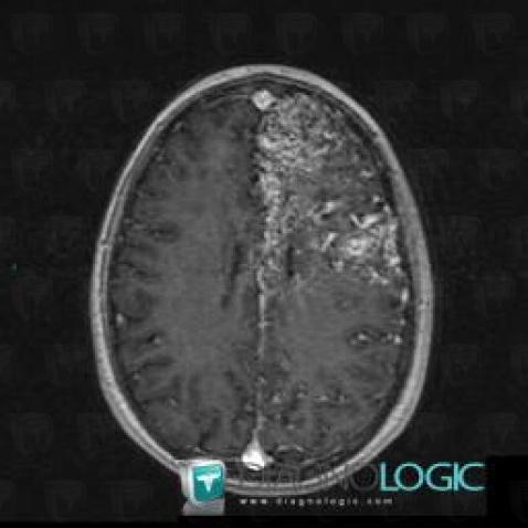
Here is the specific information in the key image above:
- Diagnosis Arteriovenous malformation, Location(s) Cortico subcortical region, with gamuts Gyriform anomalyCerebral hemispheres, with gamuts Intracerebral lesion with intense enhancement, Multifocal intracranial lesions



 Patient details - Key images