The images below illustrate this case for diagnoses Tuberculosis (link to Abscess), Tuberculosis (link to Lymphadenopathy), Tuberculosis (link to Spondylodiscitis), Tuberculosis, for the modalities (CT)
Gamuts for this case : :
- Low attenuation mediastinal mass
- Cystic mediastinal mass
- Middle mediastinal mass
- Unilateral adrenal mass
- Without gamut
- Extradural lesion
- Lesion in the psoas space
- Paraspinal soft tissue mass
- Low density retroperitoneal mass
- Cystic retroperitoneal mass
- Hematogenous / Miliary micronodules
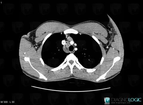
Here is the specific information in the key image above:
- Diagnosis Tuberculosis, Location(s) Mediastinum, with gamuts Low attenuation mediastinal mass, Cystic mediastinal mass, Middle mediastinal mass
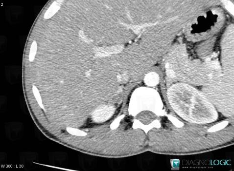
Here is the specific information in the key image above:
- Diagnosis Tuberculosis, Location(s) Adrenal glands, with gamuts Unilateral adrenal mass
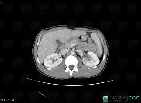
Here is the specific information in the key image above:
- Diagnosis Tuberculosis, Location(s) Collecting system, with gamuts
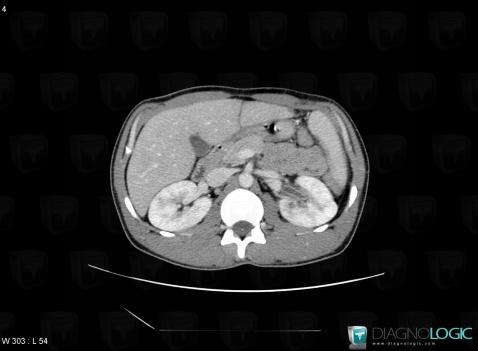
Here is the specific information in the key image above:
- Diagnosis Tuberculosis, Location(s) Collecting system, with gamuts
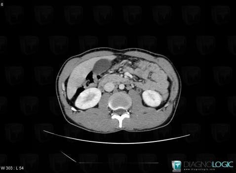
Here is the specific information in the key image above:
- Diagnosis Tuberculosis, Location(s) Ureter, with gamuts
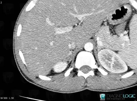
Here is the specific information in the key image above:
- Diagnosis Tuberculosis, Location(s) Adrenal glands, with gamuts Unilateral adrenal mass
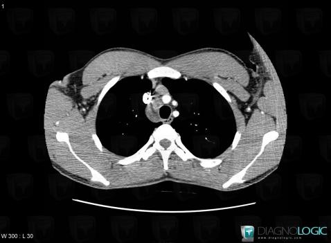
Here is the specific information in the key image above:
- Diagnosis Tuberculosis, Location(s) Mediastinum, with gamuts Low attenuation mediastinal mass, Cystic mediastinal mass, Middle mediastinal mass
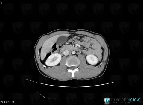
Here is the specific information in the key image above:
- Diagnosis Tuberculosis, Location(s) Ureter, with gamuts
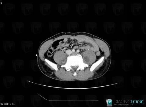
Here is the specific information in the key image above:
- Diagnosis Tuberculosis (link to Abscess), Location(s) Spinal canal / Cord, with gamuts Extradural lesion
- Diagnosis Tuberculosis, Location(s) Retroperitoneum, with gamuts Lesion in the psoas spaceVertebral body / Disk, with gamuts
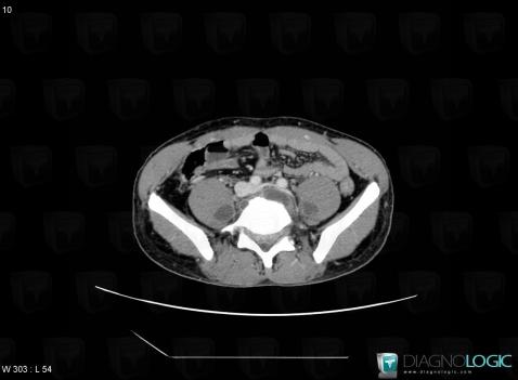
Here is the specific information in the key image above:
- Diagnosis Tuberculosis (link to Spondylodiscitis), Location(s) Paraspinal, with gamuts Paraspinal soft tissue mass
- Diagnosis Tuberculosis (link to Abscess), Location(s) Retroperitoneum, with gamuts Low density retroperitoneal mass
- Diagnosis Tuberculosis (link to Lymphadenopathy), Location(s) Retroperitoneum, with gamuts Cystic retroperitoneal mass

Here is the specific information in the key image above:
- Diagnosis Tuberculosis, Location(s) Seminal vesicles, with gamuts
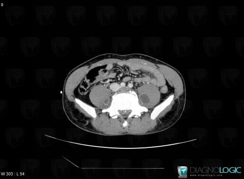
Here is the specific information in the key image above:
- Diagnosis Tuberculosis (link to Abscess), Location(s) Spinal canal / Cord, with gamuts Extradural lesion
- Diagnosis Tuberculosis, Location(s) Retroperitoneum, with gamuts Lesion in the psoas spaceVertebral body / Disk, with gamuts
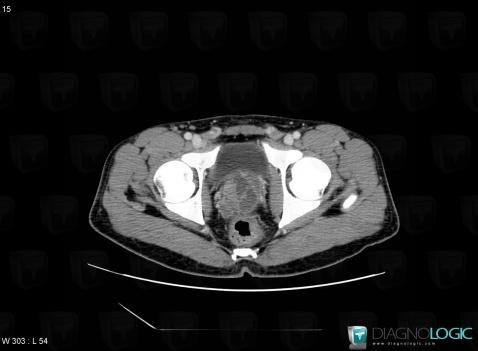
Here is the specific information in the key image above:
- Diagnosis Tuberculosis, Location(s) Seminal vesicles, with gamuts
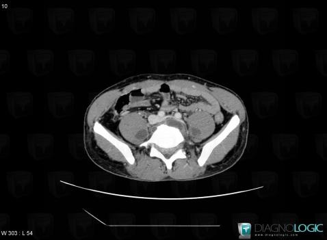
Here is the specific information in the key image above:
- Diagnosis Tuberculosis (link to Spondylodiscitis), Location(s) Paraspinal, with gamuts Paraspinal soft tissue mass
- Diagnosis Tuberculosis (link to Abscess), Location(s) Retroperitoneum, with gamuts Low density retroperitoneal mass
- Diagnosis Tuberculosis (link to Lymphadenopathy), Location(s) Retroperitoneum, with gamuts Cystic retroperitoneal mass
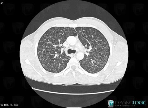
Here is the specific information in the key image above:
- Diagnosis Tuberculosis, Location(s) Pulmonary parenchyma, with gamuts Hematogenous / Miliary micronodules
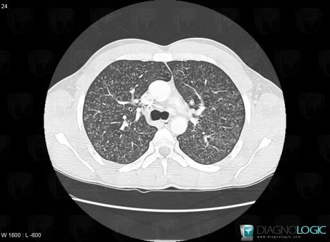
Here is the specific information in the key image above:
- Diagnosis Tuberculosis, Location(s) Pulmonary parenchyma, with gamuts Hematogenous / Miliary micronodules