The images below illustrate this case for diagnoses Tuberculosis (link to Abscess), Tuberculosis (link to Lymphadenopathy), Tuberculosis (link to Osteomyelitis), Tuberculosis (link to Pulmonary fibrosis), Tuberculosis, for the modalities (US ,X rays ,CT ,MRI)
Gamuts for this case : :
- Porta hepatis lesion
- Acute consolidation
- Upper lung zone disease
- Bilateral hilar enlargement
- Hilar lymph nodes enlargement in X ray
- Hilar mass
- Localized consolidation
- Superior mediastinal mass
- Cystic mediastinal mass
- Anterior mediastinal mass
- Cervical lymph node enlargement
- Paralaryngeal space lesion
- Cystic cervical mass
- Carotid space lesion
- Hilar lymph nodes enlargement
- Low attenuation mediastinal mass
- Middle mediastinal mass
- Posterior mediastinal mass
- Centrilobular micronodules
- Intracerebral T2W or FLAIR hyperintense lesion
- Intracerebral lesion with moderate enhancement
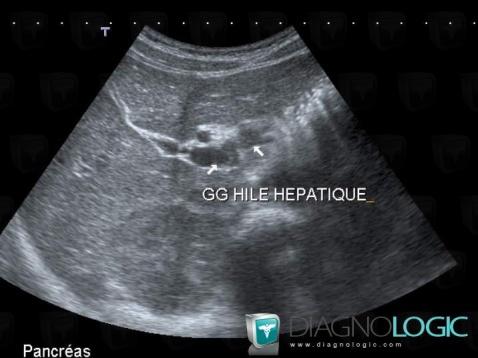
Here is the specific information in the key image above:
- Diagnosis Tuberculosis (link to Lymphadenopathy), Location(s) Liver, with gamuts Porta hepatis lesion
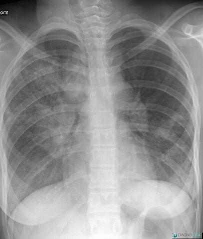
Here is the specific information in the key image above:
- Diagnosis Tuberculosis, Location(s) Pulmonary parenchyma, with gamuts Acute consolidation, Upper lung zone disease, Localized consolidationHila, with gamuts Bilateral hilar enlargement, Hilar lymph nodes enlargement in X ray
- Diagnosis Tuberculosis (link to Pulmonary fibrosis), Location(s) Hila, with gamuts Hilar mass
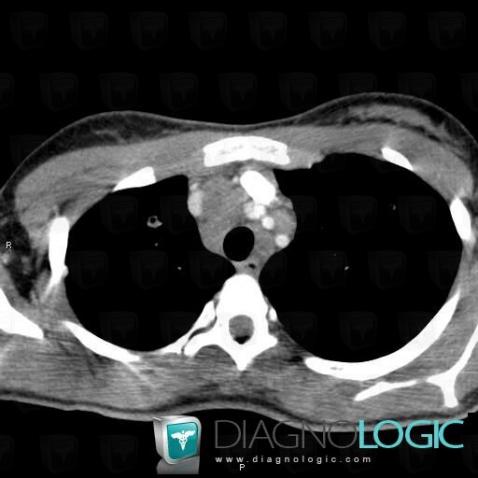
Here is the specific information in the key image above:
- Diagnosis Tuberculosis, Location(s) Mediastinum, with gamuts Superior mediastinal mass, Cystic mediastinal mass, Anterior mediastinal mass
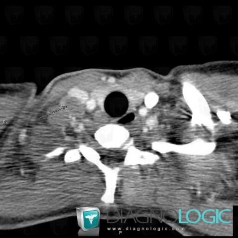
Here is the specific information in the key image above:
- Diagnosis Tuberculosis, Location(s) Deep neck spaces, with gamuts Cervical lymph node enlargement
- Diagnosis Tuberculosis (link to Lymphadenopathy), Location(s) Deep neck spaces, with gamuts Paralaryngeal space lesion, Cystic cervical mass, Carotid space lesion
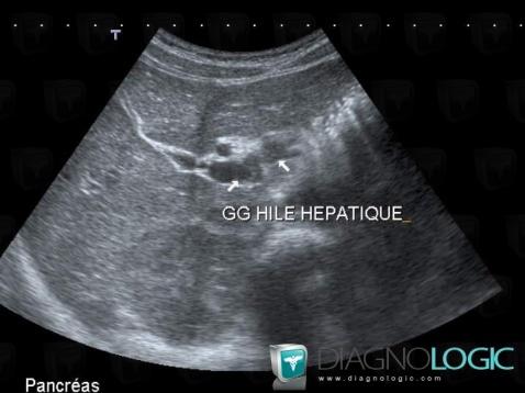
Here is the specific information in the key image above:
- Diagnosis Tuberculosis (link to Lymphadenopathy), Location(s) Liver, with gamuts Porta hepatis lesion
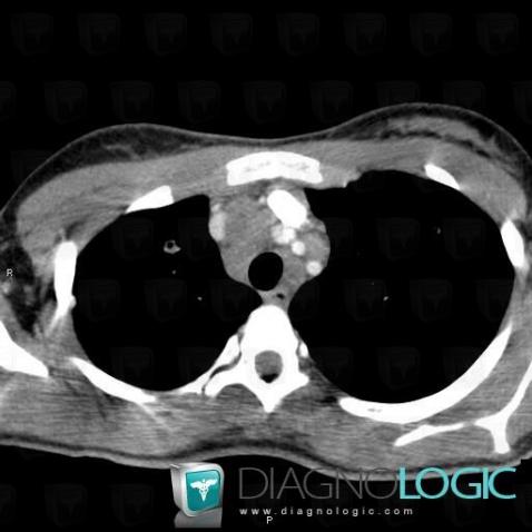
Here is the specific information in the key image above:
- Diagnosis Tuberculosis, Location(s) Mediastinum, with gamuts Superior mediastinal mass, Cystic mediastinal mass, Anterior mediastinal mass
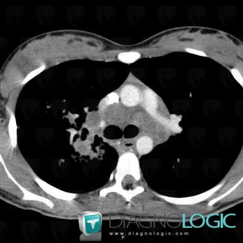
Here is the specific information in the key image above:
- Diagnosis Tuberculosis, Location(s) Mediastinum, with gamuts Hilar lymph nodes enlargement, Low attenuation mediastinal mass, Middle mediastinal mass
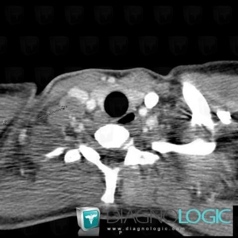
Here is the specific information in the key image above:
- Diagnosis Tuberculosis, Location(s) Deep neck spaces, with gamuts Cervical lymph node enlargement
- Diagnosis Tuberculosis (link to Lymphadenopathy), Location(s) Deep neck spaces, with gamuts Paralaryngeal space lesion, Cystic cervical mass, Carotid space lesion
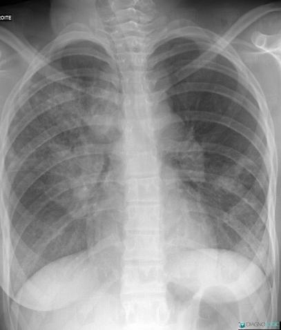
Here is the specific information in the key image above:
- Diagnosis Tuberculosis, Location(s) Pulmonary parenchyma, with gamuts Acute consolidation, Upper lung zone disease, Localized consolidationHila, with gamuts Bilateral hilar enlargement, Hilar lymph nodes enlargement in X ray
- Diagnosis Tuberculosis (link to Pulmonary fibrosis), Location(s) Hila, with gamuts Hilar mass
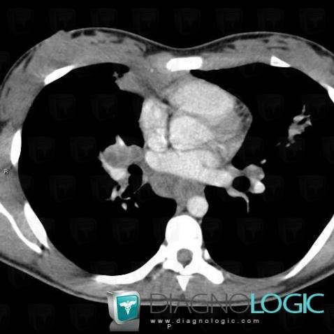
Here is the specific information in the key image above:
- Diagnosis Tuberculosis (link to Osteomyelitis), Location(s) Mediastinum, with gamuts Posterior mediastinal mass
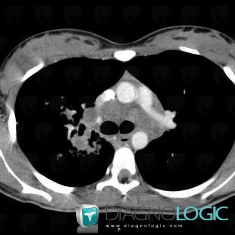
Here is the specific information in the key image above:
- Diagnosis Tuberculosis, Location(s) Mediastinum, with gamuts Hilar lymph nodes enlargement, Low attenuation mediastinal mass, Middle mediastinal mass
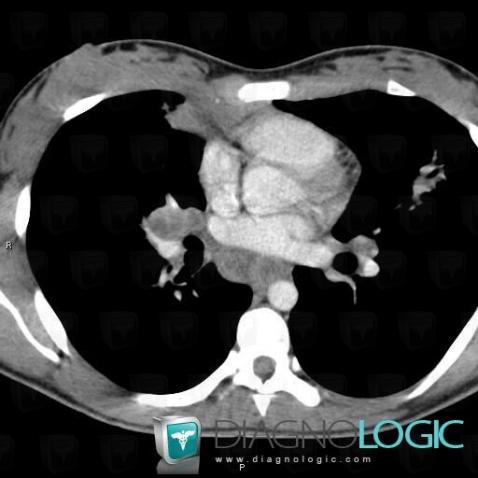
Here is the specific information in the key image above:
- Diagnosis Tuberculosis (link to Osteomyelitis), Location(s) Mediastinum, with gamuts Posterior mediastinal mass
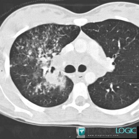
Here is the specific information in the key image above:
- Diagnosis Tuberculosis, Location(s) Pulmonary parenchyma, with gamuts Centrilobular micronodules
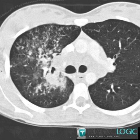
Here is the specific information in the key image above:
- Diagnosis Tuberculosis, Location(s) Pulmonary parenchyma, with gamuts Centrilobular micronodules
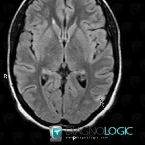
Here is the specific information in the key image above:
- Diagnosis Tuberculosis (link to Abscess), Location(s) Cerebral hemispheres, with gamuts Intracerebral T2W or FLAIR hyperintense lesion
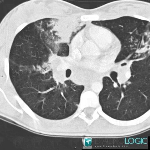
Here is the specific information in the key image above:
- Diagnosis Tuberculosis, Location(s) Pulmonary parenchyma, with gamuts Localized consolidation, Acute consolidation
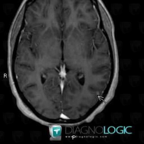
Here is the specific information in the key image above:
- Diagnosis Tuberculosis (link to Abscess), Location(s) Cerebral hemispheres, with gamuts Intracerebral lesion with moderate enhancement
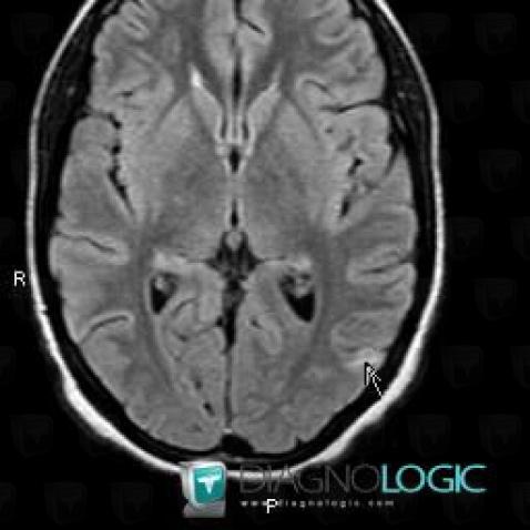
Here is the specific information in the key image above:
- Diagnosis Tuberculosis (link to Abscess), Location(s) Cerebral hemispheres, with gamuts Intracerebral T2W or FLAIR hyperintense lesion
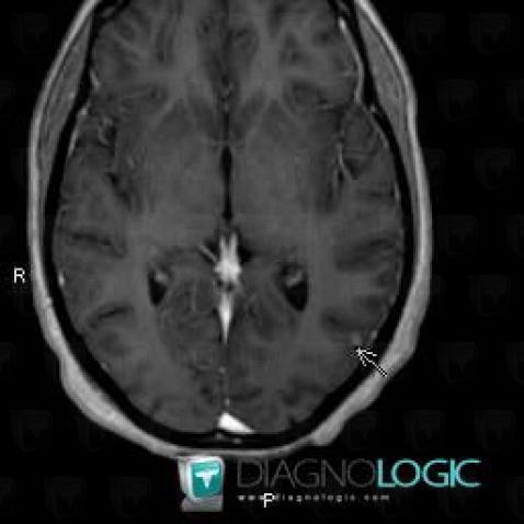
Here is the specific information in the key image above:
- Diagnosis Tuberculosis (link to Abscess), Location(s) Cerebral hemispheres, with gamuts Intracerebral lesion with moderate enhancement
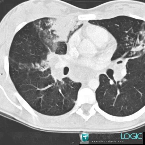
Here is the specific information in the key image above:
- Diagnosis Tuberculosis, Location(s) Pulmonary parenchyma, with gamuts Localized consolidation, Acute consolidation