The summary section allows to see, in one glance, the key features of a diagnosis
Gender :
all
Age :
5 year (Min), 75 year (Max), 20 year (Mean)
Osteoblastoma
Osteogenic benign tumor whose histological appearance is very similar to that of osteoid osteoma. The osteoid is arranged less regularly.
In borderline cases it is considered that this is a Osteoblastoma if the tumor is> 15mm
Rarer than osteoid osteoma (3.5% of benign primary tumors).
Men> Women (2/1)
Age: 5-25 years (85% of cases).
Location:
- Spine (60%), especially on the rear> femur (10%)> Tibia (8%)> Sacrum bow mandibles
Clinic:
- Very close to that of osteoid osteoma.
- Toxic form (rare): fever, poor general condition, biological inflammation
- Form locally aggressive (rare, especially sacrum).
- Spine: Painful scoliosis can
Biology
- Osteomalacia / rickets oncogenic: the tumor can secrete a hormone
- Phosphaturiante (phosphatonin or FGF 23).
Radiology / Scanner:
- Variable size and highly polymorphic radiological appearance
- Four main kinds radiological
1 Big osteoid osteoma (> 15mm)
Reaction Osteosclerosis less frequent than in osteoid osteoma multifocal mineralized matrix and noncentral
2 Injury blower near the cyst aneurysmal
The presence of intratumoral ossification (very inconsistent) may help differentiate osteoblastoma aneurysmal bone cyst.There are forms with aneurysmal bone cyst (secondary) associated with osteoblastoma.
3 osteolytic lesion pseudométastatique
4 Injury osteogenic osteosarcoma recalling with a less aggressive appearance.
- Spine: may affect two adjacent vertebrae (joint).
MRI:
- Central lesion intermediate signal in T1 and T2, taking intense contrast in the arterial phase (such as osteoid osteoma).
- Bone edema and / or adjacent soft tissues as in osteoid osteoma
- Levels sero-hematic possible especially in forms or blowers associated with aneurysmal cyst
Bone scintigraphy
- Intense uptake at the early time
Treatment:
- Careful surgical excision Reduced to curettage for sensitive locations
- Treatment by percutaneous radiofrequency or cryotherapy can be tried in the forms not too large
Frequent recurrence (30-50%)
Remember:
Highly polymorphic Image
A mention to a very limited fan lytic lesion of the posterior arch
Search for a calcified matrix for differentiating an aneurysmal cyst
Osteoblastoma


Osteogenic benign tumor whose histological appearance is very similar to that of osteoid osteoma. The osteoid is arranged less regularly.
In borderline cases it is considered that this is a Osteoblastoma if the tumor is> 15mm
Rarer than osteoid osteoma (3.5% of benign primary tumors).
Men> Women (2/1)
Age: 5-25 years (85% of cases).
Location:
- Spine (60%), especially on the rear> femur (10%)> Tibia (8%)> Sacrum bow mandibles
Clinic:
- Very close to that of osteoid osteoma.
- Toxic form (rare): fever, poor general condition, biological inflammation
- Form locally aggressive (rare, especially sacrum).
- Spine: Painful scoliosis can
Biology
- Osteomalacia / rickets oncogenic: the tumor can secrete a hormone
- Phosphaturiante (phosphatonin or FGF 23).
Radiology / Scanner:
- Variable size and highly polymorphic radiological appearance
- Four main kinds radiological
1 Big osteoid osteoma (> 15mm)
Reaction Osteosclerosis less frequent than in osteoid osteoma
multifocal mineralized matrix and noncentral
2 Injury blower near the cyst aneurysmal
The presence of intratumoral ossification (very inconsistent) may help differentiate osteoblastoma aneurysmal bone cyst. There are forms with aneurysmal bone cyst (secondary) associated with osteoblastoma.
3 osteolytic lesion pseudométastatique
4 Injury osteogenic osteosarcoma recalling with a less aggressive appearance.
- Spine: may affect two adjacent vertebrae (joint).
MRI:
- Central lesion intermediate signal in T1 and T2, taking intense contrast in the arterial phase (such as osteoid osteoma).
- Bone edema and / or adjacent soft tissues as in osteoid osteoma
- Levels sero-hematic possible especially in forms or blowers associated with aneurysmal cyst
Bone scintigraphy
- Intense uptake at the early time
Treatment:
- Careful surgical excision Reduced to curettage for sensitive locations
- Treatment by percutaneous radiofrequency or cryotherapy can be tried in the forms not too large
Frequent recurrence (30-50%)
Remember:
Highly polymorphic Image
A mention to a very limited fan lytic lesion of the posterior arch
Search for a calcified matrix for differentiating an aneurysmal cyst
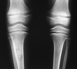
Form with oncogenic osteomalacia
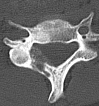
04 osteoblastoma peudométastatique blade C5. Rate aspect blowing pseudotumoral and low peripheral osteosclerosis
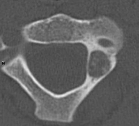
05 osteoblastoma mimicking a large cervical osteoid osteoma. Note the multifocal mineralized matrix and not of central Osteoblastoma
Tibial osteoblastoma with oncogenic hypophosphatemic rickets. Note the expansion of the growth plate associated with rickets.