The images below illustrate this case for diagnoses PVNS, for the modalities (MRI)
Gamuts for this case : :
- Periarticular T1 and T2 hypointense lesion
- Hypointense T2 WI soft tissue lesion
- Without gamut
- Synovial mass or thickening
- Soft tissue mass about a joint
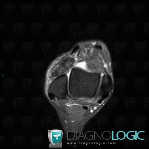
Here is the specific information in the key image above:
- Diagnosis PVNS, Location(s) Tibio/fibulo talar joint, with gamuts Periarticular T1 and T2 hypointense lesionOther soft tissues/nerves - Ankle, with gamuts Hypointense T2 WI soft tissue lesion
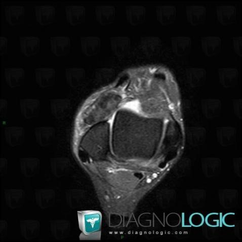
Here is the specific information in the key image above:
- Diagnosis PVNS, Location(s) Tibio/fibulo talar joint, with gamuts Periarticular T1 and T2 hypointense lesionOther soft tissues/nerves - Ankle, with gamuts Hypointense T2 WI soft tissue lesion
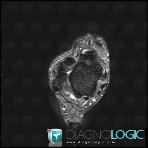
Here is the specific information in the key image above:
- Diagnosis PVNS, Location(s) Tibio/fibulo talar joint, with gamuts
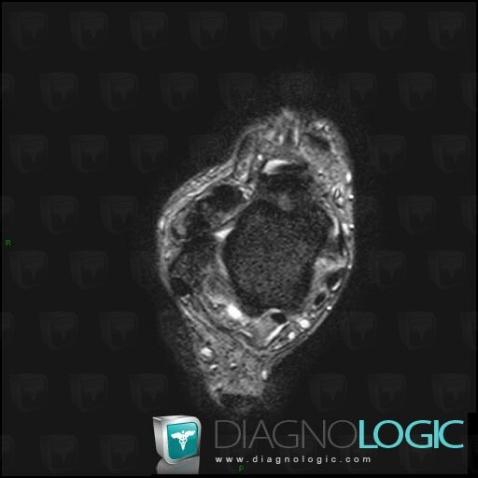
Here is the specific information in the key image above:
- Diagnosis PVNS, Location(s) Tibio/fibulo talar joint, with gamuts
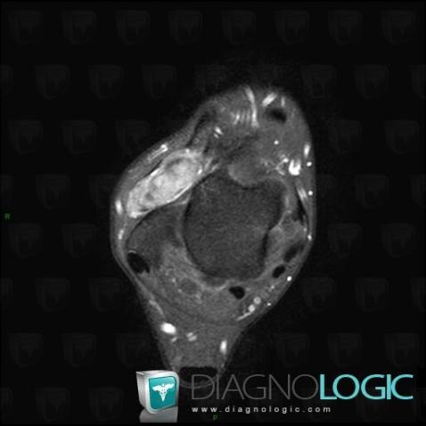
Here is the specific information in the key image above:
- Diagnosis PVNS, Location(s) Tibio/fibulo talar joint, with gamuts Synovial mass or thickening, Soft tissue mass about a joint
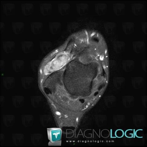
Here is the specific information in the key image above:
- Diagnosis PVNS, Location(s) Tibio/fibulo talar joint, with gamuts Synovial mass or thickening, Soft tissue mass about a joint
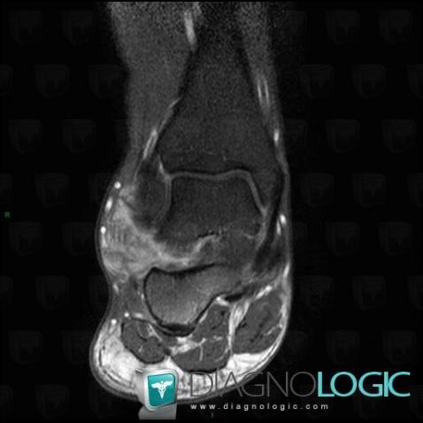
Here is the specific information in the key image above:
- Diagnosis PVNS, Location(s) Other soft tissues/nerves - Ankle, with gamuts Soft tissue mass about a joint
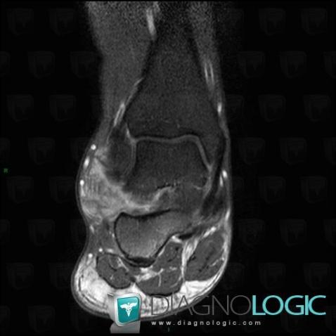
Here is the specific information in the key image above:
- Diagnosis PVNS, Location(s) Other soft tissues/nerves - Ankle, with gamuts Soft tissue mass about a joint