The images below illustrate this case for diagnoses Lipoid pneumonia, Pericardial effusion, Aortic dissection, for the modalities (CT)
Gamuts for this case : :
- Pulmonary mass with hilar attachment
- Chronic consolidation
- Localized consolidation
- Lower lung zone disease
- Solitary pulmonary mass
- thoracic vascular disease
- Without gamut
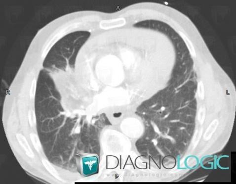
Here is the specific information in the key image above:
- Diagnosis Lipoid pneumonia, Location(s) Pulmonary parenchyma, with gamuts Pulmonary mass with hilar attachment, Chronic consolidation, Localized consolidation, Lower lung zone disease, Solitary pulmonary mass
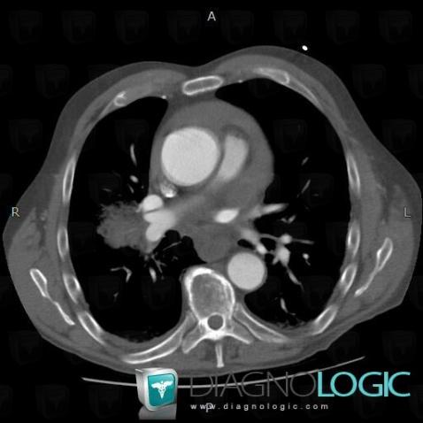
Here is the specific information in the key image above:
- Diagnosis Aortic dissection, Location(s) Aorta, with gamuts thoracic vascular disease
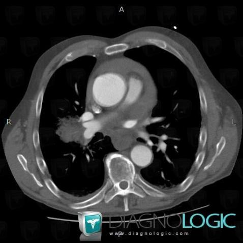
Here is the specific information in the key image above:
- Diagnosis Aortic dissection, Location(s) Aorta, with gamuts thoracic vascular disease
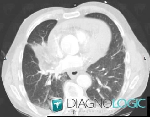
Here is the specific information in the key image above:
- Diagnosis Lipoid pneumonia, Location(s) Pulmonary parenchyma, with gamuts Pulmonary mass with hilar attachment, Chronic consolidation, Localized consolidation, Lower lung zone disease, Solitary pulmonary mass
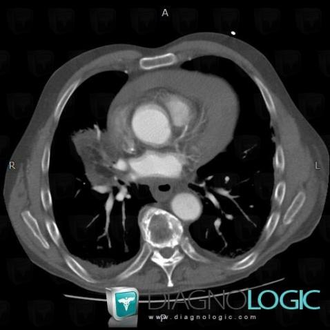
Here is the specific information in the key image above:
- Diagnosis Lipoid pneumonia, Location(s) Pulmonary parenchyma, with gamuts
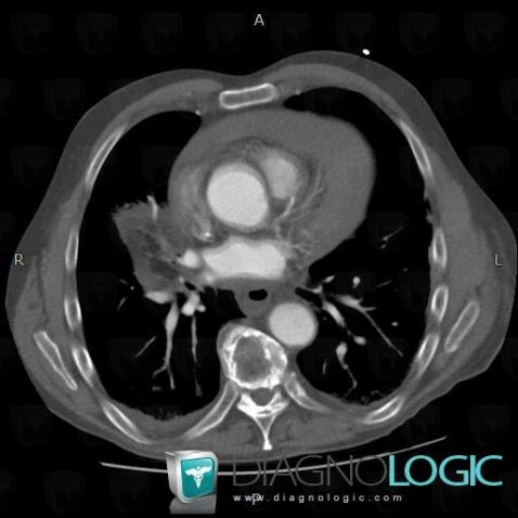
Here is the specific information in the key image above:
- Diagnosis Lipoid pneumonia, Location(s) Pulmonary parenchyma, with gamuts
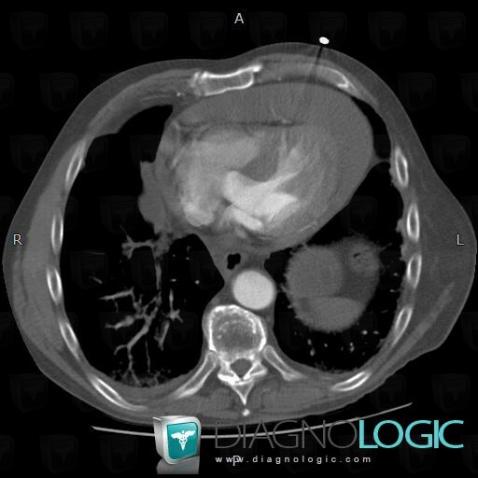
Here is the specific information in the key image above:
- Diagnosis Pericardial effusion, Location(s) Cardiac cavities / Pericardium, with gamuts
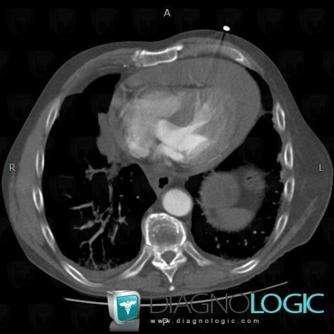
Here is the specific information in the key image above:
- Diagnosis Pericardial effusion, Location(s) Cardiac cavities / Pericardium, with gamuts