The images below illustrate this case for diagnoses Acute eosinophilic pneumonia (link to Eosinophilic pneumonia), Acute eosinophilic pneumonia, for the modalities (CT ,X rays)
Gamuts for this case : :
- Smooth peribronchovacular / interlobular thickening
- Peribronchovacular / interlobular thickening
- Disseminated consolidation
- Acute consolidation
- Localized consolidation
- Focal area of ground glass opacity
- Large symetric area of ground glass opacity
- Without gamut
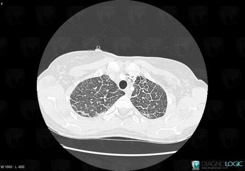
Here is the specific information in the key image above:
- Diagnosis Acute eosinophilic pneumonia, Location(s) Pulmonary parenchyma, with gamuts Smooth peribronchovacular / interlobular thickening, Peribronchovacular / interlobular thickening
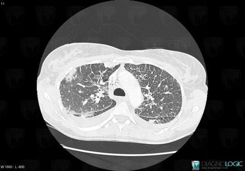
Here is the specific information in the key image above:
- Diagnosis Acute eosinophilic pneumonia (link to Eosinophilic pneumonia), Location(s) Pulmonary parenchyma, with gamuts Disseminated consolidation, Acute consolidation
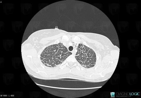
Here is the specific information in the key image above:
- Diagnosis Acute eosinophilic pneumonia, Location(s) Pulmonary parenchyma, with gamuts Smooth peribronchovacular / interlobular thickening, Peribronchovacular / interlobular thickening
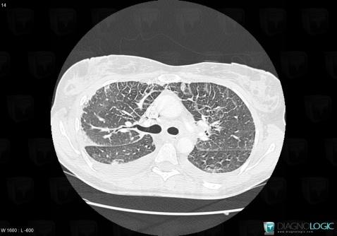
Here is the specific information in the key image above:
- Diagnosis Acute eosinophilic pneumonia (link to Eosinophilic pneumonia), Location(s) Pulmonary parenchyma, with gamuts Localized consolidation
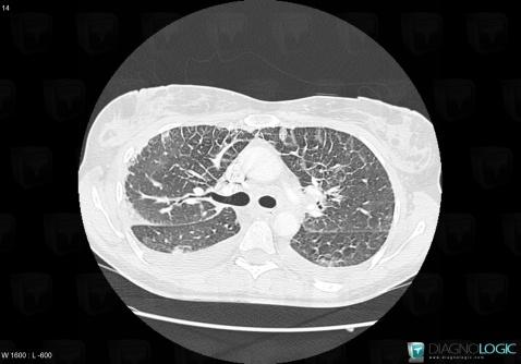
Here is the specific information in the key image above:
- Diagnosis Acute eosinophilic pneumonia (link to Eosinophilic pneumonia), Location(s) Pulmonary parenchyma, with gamuts Localized consolidation
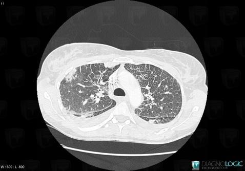
Here is the specific information in the key image above:
- Diagnosis Acute eosinophilic pneumonia (link to Eosinophilic pneumonia), Location(s) Pulmonary parenchyma, with gamuts Disseminated consolidation, Acute consolidation
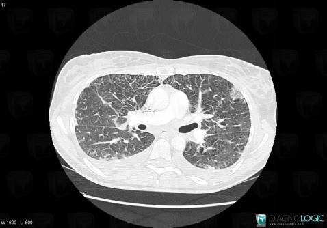
Here is the specific information in the key image above:
- Diagnosis Acute eosinophilic pneumonia (link to Eosinophilic pneumonia), Location(s) Pulmonary parenchyma, with gamuts Focal area of ground glass opacity
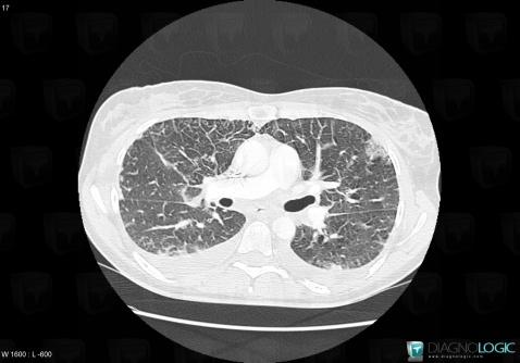
Here is the specific information in the key image above:
- Diagnosis Acute eosinophilic pneumonia (link to Eosinophilic pneumonia), Location(s) Pulmonary parenchyma, with gamuts Focal area of ground glass opacity
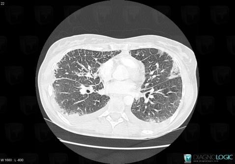
Here is the specific information in the key image above:
- Diagnosis Acute eosinophilic pneumonia (link to Eosinophilic pneumonia), Location(s) Pulmonary parenchyma, with gamuts Large symetric area of ground glass opacity
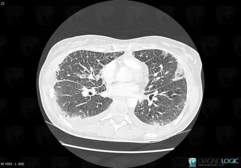
Here is the specific information in the key image above:
- Diagnosis Acute eosinophilic pneumonia (link to Eosinophilic pneumonia), Location(s) Pulmonary parenchyma, with gamuts Large symetric area of ground glass opacity
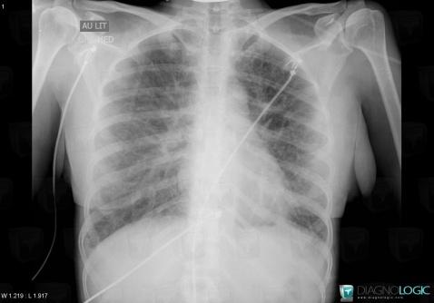
Here is the specific information in the key image above:
- Diagnosis Acute eosinophilic pneumonia, Location(s) Pulmonary parenchyma, with gamuts
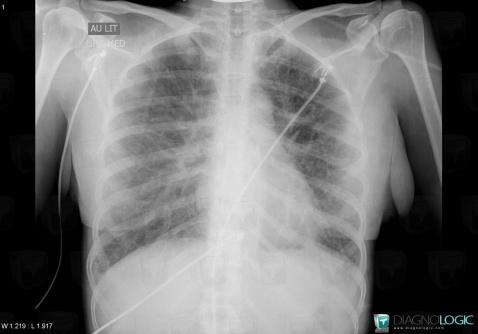
Here is the specific information in the key image above:
- Diagnosis Acute eosinophilic pneumonia, Location(s) Pulmonary parenchyma, with gamuts