The images below illustrate this case for diagnoses Colloid cyst, for the modalities (MRI ,CT)
Gamuts for this case : :
- Midline mass
- Intracerebral T1W hyperintense lesion
- Hyperdense intracerebral lesion on noncontrast CT
- Ventricular wall nodules
- Intracerebral T2W or FLAIR hypointense lesion
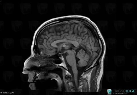
Here is the specific information in the key image above:
- Diagnosis Colloid cyst, Location(s) Cerebral falx / Midline, with gamuts Midline mass
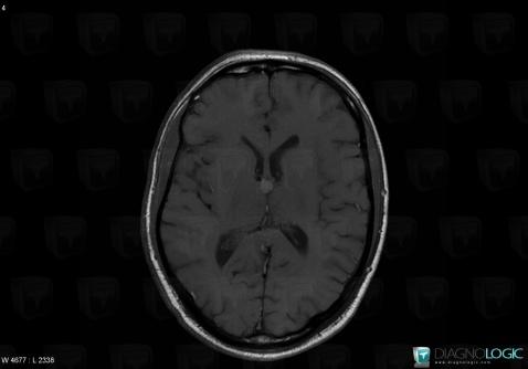
Here is the specific information in the key image above:
- Diagnosis Colloid cyst, Location(s) Cerebral hemispheres, with gamuts Intracerebral T1W hyperintense lesion
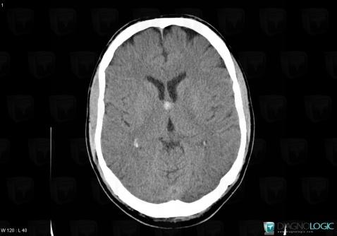
Here is the specific information in the key image above:
- Diagnosis Colloid cyst, Location(s) Cerebral hemispheres, with gamuts Hyperdense intracerebral lesion on noncontrast CTCerebral falx / Midline, with gamuts Midline massVentricles / Periventricular region, with gamuts Ventricular wall nodules
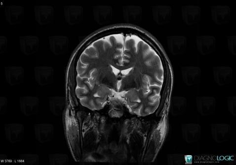
Here is the specific information in the key image above:
- Diagnosis Colloid cyst, Location(s) Cerebral hemispheres, with gamuts Intracerebral T2W or FLAIR hypointense lesion
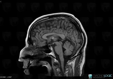
Here is the specific information in the key image above:
- Diagnosis Colloid cyst, Location(s) Cerebral falx / Midline, with gamuts Midline mass
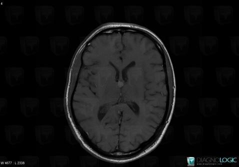
Here is the specific information in the key image above:
- Diagnosis Colloid cyst, Location(s) Cerebral hemispheres, with gamuts Intracerebral T1W hyperintense lesion
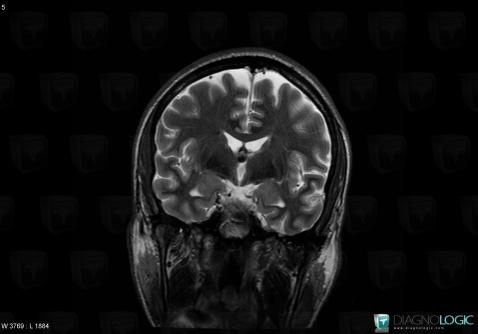
Here is the specific information in the key image above:
- Diagnosis Colloid cyst, Location(s) Cerebral hemispheres, with gamuts Intracerebral T2W or FLAIR hypointense lesion
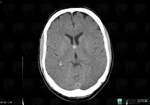
Here is the specific information in the key image above:
- Diagnosis Colloid cyst, Location(s) Cerebral hemispheres, with gamuts Hyperdense intracerebral lesion on noncontrast CTCerebral falx / Midline, with gamuts Midline massVentricles / Periventricular region, with gamuts Ventricular wall nodules