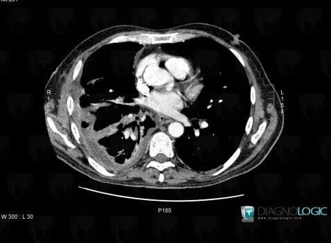
The images below illustrate this case for diagnoses Pseudomonas pneumonia (link to Abscess), Empyema, Pseudomonas pneumonia, for the modalities (CT)
Gamuts for this case : :
- Pleural thickening
- Multiples pleural masses
- Disseminated consolidation
- Cavitary pulmonary mass
- Large symetric area of ground glass opacity
- Solitary pulmonary mass
- Acute consolidation
- Localized consolidation

Here is the specific information in the key image above:
- Diagnosis Empyema, Location(s) Pleura, with gamuts Pleural thickening, Multiples pleural masses

Here is the specific information in the key image above:
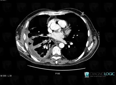
- Diagnosis Empyema, Location(s) Pleura, with gamuts Pleural thickening, Multiples pleural masses

Here is the specific information in the key image above:
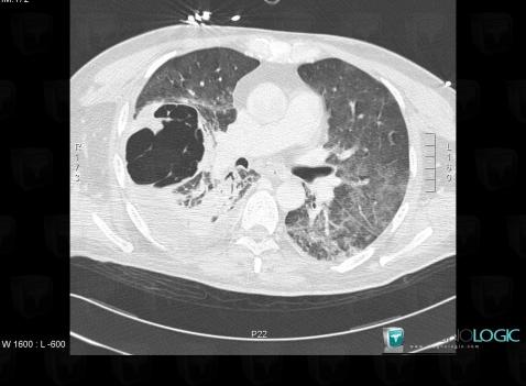
- Diagnosis Pseudomonas pneumonia, Location(s) Pulmonary parenchyma, with gamuts Disseminated consolidation, Large symetric area of ground glass opacity, Acute consolidation, Localized consolidation
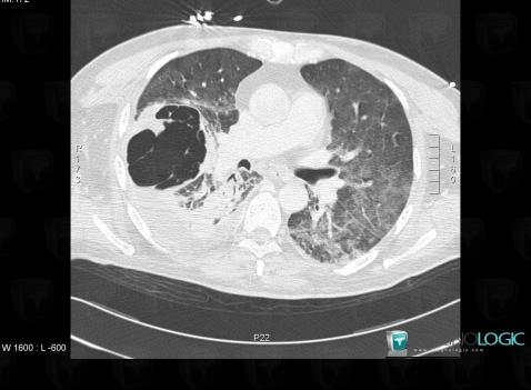
- Diagnosis Pseudomonas pneumonia (link to Abscess), Location(s) Pulmonary parenchyma, with gamuts Cavitary pulmonary mass, Solitary pulmonary mass